
Animal Tissue
Class 12 Biology Notes • NEB Syllabus 2083
Animal Tissue for Class 12 Biology Unit 6: syllabus, micro syllabus, 40 solved multiple choice questions, short and long answer questions on epithelial, connective, muscular and nervous tissue, with figures and two PDF notes.
1. Animal Tissue Syllabus
Unit 6: Animal Tissue (Chapter 1)
Animal Tissue are four: epithelial, connective, muscular and nervous tissue. A tissue is a group of cells with a common origin that work together. These notes follow the NEB Class 12 Biology syllabus (+2 Biology, Nepal), Unit 6, chapter 1. Practise the multiple choice questions, then the solved short and long answers with figures, then the PDF notes.

Figure: Animal tissue, Class 12 Biology (NEB)
Figure: Classification of the four types of animal tissue
Micro syllabus: 6. Animal Tissues
Read more about tissue, epithelium and connective tissue on Wikipedia. More notes: Class 12 Biology notes, Class 12 Chemistry notes and Class 12 Physics notes.
2. Multiple Choice Questions
Question 1. Who coined the term tissue?
- a) Grew
- b) Bichat
- c) Mayer
- d) Robert Hooke
Correct Answer: b) Bichat
Reason: Marie Francois Xavier Bichat, a French anatomist, coined ’tissue’ for animals and is called the father of histology. Mayer coined ‘histology’.
Question 2. What is meant by a tissue?
- a) Group of cells of similar origin
- b) Group of cells of dissimilar origin
- c) Group of similar or dissimilar cells
- d) Group of unrelated cells
Correct Answer: a) Group of cells of similar origin
Reason: Cells of a tissue have a common origin, though their structure, location and function may be similar or dissimilar.
Question 3. Which of the following tissue is not animal tissue?
- a) Epithelial tissue
- b) Connective tissue
- c) Parenchymatous tissue
- d) Muscular tissue
Correct Answer: c) Parenchymatous tissue
Reason: Parenchyma is a plant ground tissue. The four animal tissues are epithelial, connective, muscular and nervous.
Question 4. Which one of the following animal tissue is originated from all the three germ layers?
- a) Epithelial tissue
- b) Connective tissue
- c) Muscular tissue
- d) Nervous tissue
Correct Answer: a) Epithelial tissue
Reason: Epithelium arises from ectoderm, mesoderm and endoderm. Connective and muscular tissue are mesodermal; nervous tissue is ectodermal.
Question 5. Which one of the following statements is incorrect regarding epithelial tissue?
- a) Cells are compactly packed
- b) Provides a covering for some parts of the body
- c) Cells may have cilia
- d) It doesn’t have free border.
Correct Answer: d) It doesn’t have free border.
Reason: Epithelium always has a free (apical) border, on which cilia or microvilli may occur. The other statements are correct.
Question 6. Which one of the following is mesothelium?
- a) Tunica interna
- b) Capillary
- c) Splanchnic epithelium
- d) Endocardium
Correct Answer: c) Splanchnic epithelium
Reason: Simple squamous epithelium of mesodermal origin (parietal and visceral/splanchnic epithelium, mesenteries) is mesothelium. Tunica interna, capillary and endocardium are endothelium.
Question 7. Presence of basement membrane is the characteristic features of
- a) epithelial tissue
- b) connective tissue
- c) muscular tissue
- d) nervous tissue
Correct Answer: a) epithelial tissue
Reason: A collagen basement membrane supports the single layer of simple epithelium and the innermost layer of compound epithelium.
Question 8. The mucosal layer of intestine is made up of
- a) Stratified squamous epithelium
- b) Stratified cuboidal epithelium
- c) Stratified columnar epithelium
- d) Transitional epithelium
Correct Answer: c) Stratified columnar epithelium
Reason: Intestinal mucosa is columnar (brush-bordered simple columnar) epithelium. Only option c has columnar cells, so it is the nearest choice.
Question 9. Which of the following tissue is found in skin?
- a) Stratified squamous epithelium
- b) Stratified cuboidal epithelium
- c) Stratified columnar epithelium
- d) Pseudostratified epithelium
Correct Answer: a) Stratified squamous epithelium
Reason: Skin has keratinized stratified squamous epithelium, which is waterproof and protective.
Question 10. Which one of the following epithelium forms innermost lining of vagina?
- a) Cuboidal epithelium
- b) Columnar epithelium
- c) Stratified keratinized epithelium
- d) Stratified non-keratinized epithelium
Correct Answer: d) Stratified non-keratinized epithelium
Reason: Non-keratinized stratified squamous epithelium has a smooth surface and keeps the vagina moist.
Question 11. Plastic epithelium is
- a) striated muscle
- b) non-striated muscle
- c) urothelium
- d) cardiac muscle
Correct Answer: c) urothelium
Reason: Transitional epithelium, also called urothelium or plastic epithelium, changes shape from rounded to flat when stretched.
Question 12. Which one of the following animal tissue is most abundant?
- a) Epithelial tissue
- b) Connective tissue
- c) Muscular tissue
- d) Nervous tissue
Correct Answer: b) Connective tissue
Reason: Connective tissue is considered the most abundant animal tissue and has a large amount of matrix.
Question 13. All the three components of cells, matrix, and fibres are present in
- a) epithelial tissue
- b) connective tissue
- c) muscular tissue
- d) nervous tissue
Correct Answer: b) connective tissue
Reason: Connective tissue has three histo-components (cells, matrix, fibres). The other three have cells and minimal matrix.
Question 14. Which cells of areolar tissue are able to move and ingest foreign particles?
- a) Fibroblasts
- b) Mast cells
- c) Plasma cells
- d) Histiocytes
Correct Answer: d) Histiocytes
Reason: Histiocytes are large amoeboid macrophages that engulf foreign particles, dead cells and microbes.
Question 15. Which one of the following tissue possesses fat globules?
- a) Areolar tissue
- b) Adipose tissue
- c) Loose connective tissue
- d) Dense connective tissue
Correct Answer: b) Adipose tissue
Reason: Adipose tissue is areolar tissue modified with adipocytes, which secrete and store fat.
Question 16. Suture joints in skull bones is
- a) areolar tissue
- b) white fibrous tissue
- c) elastic connective tissue
- d) reticular tissue
Correct Answer: b) white fibrous tissue
Reason: White fibrous tissue with thick collagen bundles keeps skull bones immovable at sutures.
Question 17. Which of the following fibre is present abundantly in tendon?
- a) White collagen fibre
- b) Yellow elastic fibre
- c) Colourless reticulate fibre
- d) Red muscle fibre
Correct Answer: a) White collagen fibre
Reason: Tendon is dense connective tissue made of thin bundles of collagen fibres.
Question 18. Dense connective tissue contains tendon and ligament. If tendon connects muscle to bone, which structure is connected by ligament?
- a) Bone to bone
- b) Muscle to bone
- c) Muscle to muscle
- d) Tendon to ligament
Correct Answer: a) Bone to bone
Reason: Ligament joins bone to bone. Tendon joins bone to muscle.
Question 19. Ligaments are connective tissue. What are ligaments made up of?
- a) Predominantly white collagen fibres
- b) Predominantly yellow fibres
- c) Both white and yellow fibres
- d) Only white fibres
Correct Answer: a) Predominantly white collagen fibres
Reason: Ligament has thick bundles of collagen fibres along with some elastic fibres, so collagen predominates.
Question 20. Which one of the following is avascular?
- a) Tendon
- b) Ligament
- c) Cartilage
- d) Bone
Correct Answer: c) Cartilage
Reason: Cartilage has no blood vessels; nutrients and gases reach it by diffusion. Bone is vascular.
Question 21. Haversian system is diagnostic feature of bone. This system is present in which one of the following animal?
- a) Amphibians
- b) Reptile
- c) Aves
- d) Mammal
Correct Answer: d) Mammal
Reason: Osteons are seen in the diaphysis of mammalian long bone. Amphibian lamellae have no osteon.
Question 22. The human RBCs is
- a) spherical and biconvex
- b) spherical and biconcave
- c) rounded and biconvex
- d) rounded and biconcave
Correct Answer: d) rounded and biconcave
Reason: Mammalian RBCs are rounded, biconcave and non-nucleated, which gives more surface area for O2 binding.
Question 23. The function of erythrocytes is one of the following.
- a) Engulfing bacteria
- b) Producing antibodies
- c) Carrying oxygen
- d) Clotting blood
Correct Answer: c) Carrying oxygen
Reason: RBCs carry oxygen as oxyhaemoglobin and carbon dioxide as carbaminohaemoglobin.
Question 24. Which pair of white blood cells contains phagocytic cell?
- a) Monocytes and eosinophils
- b) Monocytes and neutrophils
- c) Eosinophils and basophils
- d) Monocytes and lymphocytes
Correct Answer: b) Monocytes and neutrophils
Reason: Neutrophils (most active) and monocytes are phagocytic. Basophils show allergy and lymphocytes make antibodies.
Question 25. Excess number of white blood corpuscles in the blood results in a condition called
- a) Down syndrome
- b) Leukemia
- c) Dengue
- d) Haemophilia
Correct Answer: b) Leukemia
Reason: Leukemia is blood cancer with a very high WBC count. Leucopenia is the opposite (fewer WBCs).
Question 26. Which one of the following myocytes of the muscles are syncytial?
- a) Striped muscles
- b) Unstriped muscles
- c) Heart muscles
- d) Smooth muscles
Correct Answer: a) Striped muscles
Reason: A striped muscle fibre is a single syncytial myocyte with many nuclei in the sarcoplasm.
Question 27. The epimysium is the covering of ……………………. of striped muscles.
- a) myofibril
- b) myofibre
- c) fascicle
- d) block
Correct Answer: d) block
Reason: Endomysium covers a myofibre, perimysium a fascicle and epimysium the whole block (muscle).
Question 28. The muscle responsible for the movement of tongue and speech is one of the followings.
- a) Striated muscle
- b) Unstriated muscle
- c) Cardiac muscle
- d) Smooth muscle
Correct Answer: a) Striated muscle
Reason: Striped muscle occurs in the tongue and oesophagus and is voluntary.
Question 29. Which one of the following muscle has no stripes?
- a) Skeletal muscle
- b) Smooth muscle
- c) Voluntary muscle
- d) Cardiac muscle
Correct Answer: b) Smooth muscle
Reason: Unstriped (smooth) muscle has spindle-shaped myocytes with no transverse stripes.
Question 30. Arrector pilli is associated with
- a) striped muscle
- b) unstriped muscle
- c) cardiac muscle
- d) striated muscle
Correct Answer: b) unstriped muscle
Reason: Arrector pili is multi-unit unstriped muscle. Blood vessels are the other example.
Question 31. Which one of the following muscle has intercalated disc?
- a) Striped muscle
- b) Unstriped muscle
- c) Cardiac muscle
- d) Voluntary muscle
Correct Answer: c) Cardiac muscle
Reason: Intercalated discs separate adjacent cardiocytes of cardiac muscle.
Question 32. In the wall of heart, there is presence of cardiac muscles. Which of the following is correct for cardiac muscle?
- a) Striped and voluntary
- b) Unstriped and voluntary
- c) Striped and involuntary
- d) Unstriped and involuntary
Correct Answer: c) Striped and involuntary
Reason: Cardiac muscle has faint transverse stripes, works involuntarily and never fatigues.
Question 33. Which one of the following tissue connect neurones?
- a) Areolar tissue
- b) White fibrous tissue
- c) Reticular tissue
- d) Neuroglia
Correct Answer: d) Neuroglia
Reason: Proper connective tissue never binds nervous tissue. Neurones are bound by neuroglia.
Question 34. Which one of the following animal tissue perform no mitosis?
- a) Epithelial tissue
- b) Connective tissue
- c) Muscular tissue
- d) Nervous tissue
Correct Answer: d) Nervous tissue
Reason: Neurones cannot divide, so they cannot regenerate (neuroglia can divide).
Question 35. Which one of the following is macrophage in CNS?
- a) Histiocytes
- b) Microglia
- c) Dust cells
- d) Langerhans cells
Correct Answer: b) Microglia
Reason: Microglia devour bacteria and dead neurones in the brain. Histiocytes are in connective tissue, dust cells in lungs, Langerhans cells in skin.
Question 36. In which part of the neurone, do you find Nissl’s granules?
- a) Cyton
- b) Axon
- c) Axon terminal
- d) All
Correct Answer: a) Cyton
Reason: Nissl’s granules (RNA, ribosomes, RER) lie in the soma (cyton) and are absent at the axon hillock.
Question 37. The bipolar neurones are found in
- a) brain
- b) spinal cord
- c) retina
- d) dorsal root ganglia
Correct Answer: c) retina
Reason: A bipolar neurone has two neurites in opposite directions, as in the retina. Dorsal root ganglia have unipolar/pseudopolar neurones.
Question 38. The gray matter is found in
- a) outer part of brain and spinal cord
- b) inner part of brain and spinal cord
- c) outer part of brain and inner part of spinal cord
- d) inner part of brain and outer part of spinal cord
Correct Answer: c) outer part of brain and inner part of spinal cord
Reason: Gray matter is outer in the brain and inner in the spinal cord. White matter is the reverse.
Question 39. Which one of the following cells produce cerebrospinal fluid?
- a) Neurosecretory cells
- b) Ependymal cells
- c) Goblet cells
- d) Mast cells
Correct Answer: b) Ependymal cells
Reason: Ependymal cells line the ventricles of the brain and secrete CSF.
Question 40. Which one of the following is longest animal tissue?
- a) Epithelial tissue
- b) Connective tissue
- c) Muscular tissue
- d) Nervous tissue
Correct Answer: d) Nervous tissue
Reason: Neurones, with long axons, are the longest animal tissue.
3. Short and Long Answer Questions
Animal Tissue: Short Answer Questions
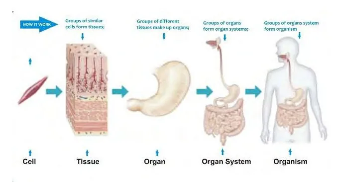
Figure: Cell, tissue, organ (stomach), organ system (digestive system) and organism
The figure shows the levels of organisation of the body, from simple to complex: cell → tissue → organ (stomach) → organ system (digestive system) → organism.
Thus tissues in animals and plants act as building blocks that form organs.
Similarities
Differences
| Simple epithelium | Compound epithelium |
|---|---|
| Single layer of cells. | Many layers of cells. |
| All cells rest on the basement membrane. | Only the innermost layer rests on it; the topmost layer has the free border. |
| Squamous, cuboidal, columnar and pseudostratified types. | Stratified (squamous, cuboidal, columnar) and transitional types. |
| For absorption, secretion, filtration and diffusion. | For protection against stress, wear and tension. |
| Found in alveoli, kidney tubules, intestine. | Found in skin, buccal cavity, urinary bladder. |
Yes. Tessellated epithelium is another name for simple squamous epithelium, also called pavement or mosaic epithelium. Its thin, flat, polygonal cells fit together like floor tiles.
Difference
| Feature | Tessellated (squamous) | Simple cuboidal | Simple columnar |
|---|---|---|---|
| Cell shape | Thin, flattened, polygonal | Cube-like, equal height and width | Tall, cylindrical, height more than width |
| Nucleus | Flattened, oval | Large, spherical, central | Elongated, oval, basal |
| Location | Alveoli, Bowman’s capsule, capillary | Kidney tubules, thyroid vesicles, ducts | Stomach, intestine, gall bladder |
| Function | Gaseous exchange, filtration | Secretion, absorption | Secretion, absorption, excretion |
Figure: Transitional epithelium (plastic epithelium) in relaxed and stretched states
Yes. Transitional epithelium is also called plastic epithelium or urothelium. It is a compound epithelium that bears stress or tension.
Figure: Types of connective tissue: proper, supportive and fluid
Connective tissue binds, supports or circulates. It is mesodermal, the most abundant animal tissue, and has fewer cells, abundant matrix and abundant fibres.
Types and functions
| Type | Examples | Function |
|---|---|---|
| Proper | Areolar, adipose, white fibrous, tendon, ligament, elastic, reticular | Binds similar and dissimilar tissues (never nervous tissue). |
| Supportive | Cartilage and bone | Supports the animal by forming the framework or endoskeleton. |
| Fluid | Blood and lymph | Transports materials, regulates the body and protects against disease. |
Areolar tissue is the soft, loose-matrix connective tissue of proper connective tissue. Cells and fibres lie in a jelly-like matrix.
Figure: Haversian system (osteon) in the compact bone of a mammal, transverse section
The Haversian system or osteon is seen in the compact bone (diaphysis) of mammalian long bone. It is absent in epiphysis and in amphibian bone.
Formed elements (blood cells or corpuscles) are the solid part of blood, about 45% of its volume. Plasma forms 55%. They are formed by haemopoiesis in red bone marrow, spleen, lymph nodes and thymus.
Types
| Formed element | Features | Function |
|---|---|---|
| RBCs (erythrocytes) | Biconcave, non-nucleated in mammals, with haemoglobin; 5 million mm-3 in male; life 120 days. | Carry O2 and CO2. |
| WBCs (leucocytes) | Granulocytes (neutrophils, basophils, eosinophils) and agranulocytes (lymphocytes, monocytes); 4500-10,000 mm-3. | Phagocytosis, allergy and antibody formation. |
| Platelets (thrombocytes) | Colourless cell fragments in mammals; 0.15-0.4 million mm-3; life 7-10 days. | Release thromboplastin for blood clotting. |
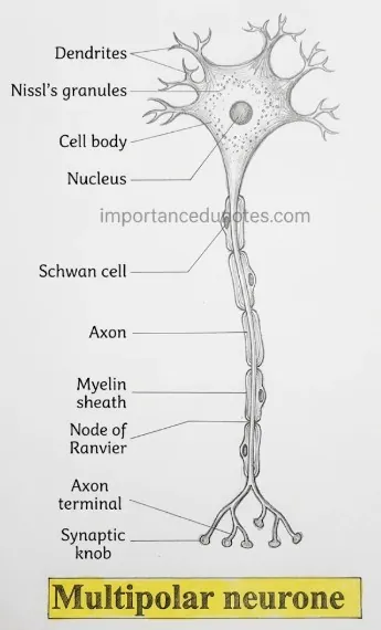
Figure: Multipolar neurone
Animal Tissue: Long Answer Questions
Simple epithelium has a single layer of cells resting on a basement membrane. On the basis of cell shape it has four types.
1. Simple squamous epithelium
Thin, flattened, polygonal cells with a flat, oval nucleus (tessellated epithelium). Location and function: alveoli (gaseous exchange), Bowman’s capsule (ultrafiltration), coelom and viscera (lining and covering), descending limb of loop of Henle (reabsorption), capillary (blood flow). Endothelium lines capillary, endocardium and tunica interna; mesothelium forms the parietal and visceral epithelium and mesenteries.
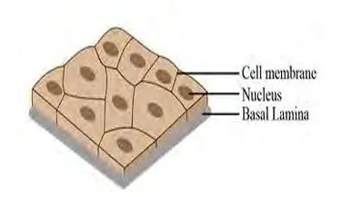
Figure: Simple squamous epithelium
2. Simple cuboidal epithelium
Cube-like cells with a large, spherical, central nucleus. It is non-ciliated (ovary, thyroid vesicles, kidney tubules, salivary and pancreatic ducts), ciliated (respiratory bronchioles, to filter air) or brush-bordered (PCT of nephron, for reabsorption).
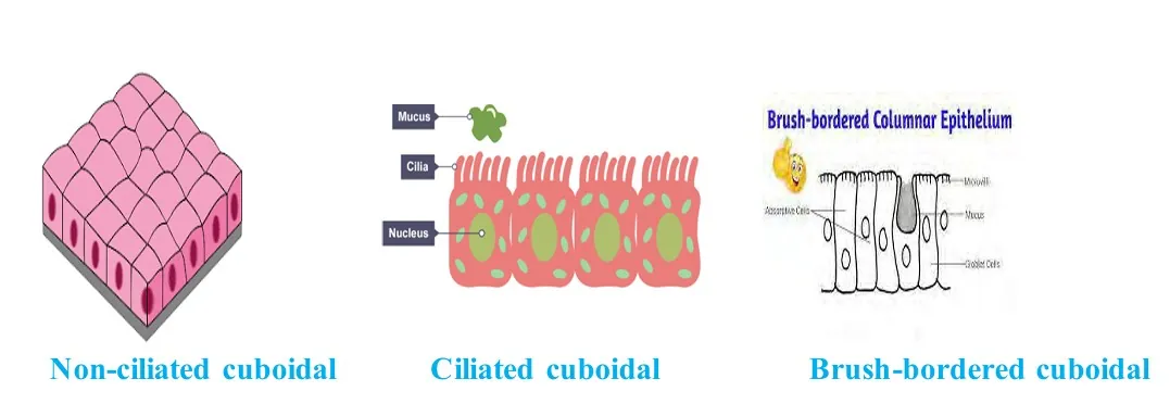
Figure: Simple cuboidal epithelium
3. Simple columnar epithelium
Tall, cylindrical cells with an elongated, oval, basal nucleus. It is non-ciliated (gall bladder, stomach, intestine), ciliated (fallopian tube, oviduct, neurocoel) or brush-bordered (intestinal mucosa, for absorption).
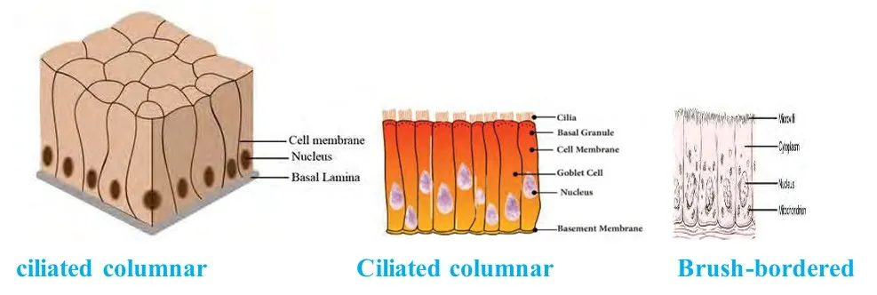
Figure: Simple columnar epithelium
4. Pseudostratified epithelium
A single layer of cuboidal and columnar cells on one basement membrane. About half of the columnar cells become goblet cells (mucus) or ciliated cells. It lines the upper respiratory tract, trachea, bronchi and bronchioles and filters dust.
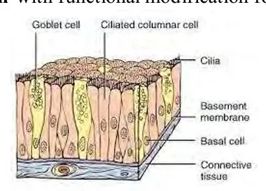
Figure: Pseudostratified epithelium
Among compound epithelia, two stratified types are less common. In both, the topmost five or six layers have the same shape and the deeper layers rest on the basement membrane. Stratified epithelium bears no stress and has a general function (protection).
Stratified cuboidal epithelium (rare)
Stratified columnar epithelium (uncommon)
Note
Stratified squamous epithelium is common: non-keratinized (buccal cavity, oesophagus, anus, vagina) and keratinized (skin and its derivatives).
In modified epithelium the free border of the cells is specialised. It has six types.
Types
| Type | Feature | Location and function |
|---|---|---|
| Ciliated | Kino-ciliated has cilia with 9+2 microtubules; stereo-ciliated has 0+0. | Respiratory organs, female reproductive organs, neurocoel (movement of mucus, ova, CSF); epididymis (sperm maturation). |
| Brush-bordered | Microvilli on the free border. | Kidney tubules and small intestine; more absorptive area. |
| Neuro-sensory | Sensory hairs joined to nerve fibres. | Olfactory epithelium; smelling. |
| Pigmented | Pigment-containing cells. | Retina. |
| Germinal | Gamete-forming cells. | Testes and ovaries. |
| Glandular | Enzyme- and hormone-secreting gland cells. | Gastric glands, pancreas, pituitary and others. |
Glandular epithelium is modified epithelium having enzyme- and hormone-secreting cells called glands.
Types of glands
| Basis | Types and examples |
|---|---|
| Ducts and secretion | Exocrine (ducted, enzymes: gastric, intestinal); endocrine (ductless, hormones: pineal, pituitary); heterocrine or mixed (pancreas). |
| Consistency of secretion | Serous (watery: sweat, tear), mucous (goblet cells) and mixed. |
| Quantity of secretion | Apocrine (pinch of cell: mammary), merocrine (little amount: salivary, pancreas), holocrine (whole cell: sebaceous). |
| Number of cells | Unicellular (goblet cell) and multicellular (sweat gland, liver, thyroid). |
| Structure | Simple (single secretory unit: tubular, branched tubular, coiled tubular, alveolar, branched alveolar) and compound (liver, salivary glands, pancreas and mammary glands). |
| Feature | Cartilage | Bone |
|---|---|---|
| Nature | Strong, flexible, avascular. | Strongest, non-flexible, vascular. |
| Cells | Chondroblasts and chondrocytes. | Osteoblasts and osteocytes. |
| Cells in lacuna | Single or in groups of 2, 3 or 4. | Single; lacuna has canaliculi. |
| Matrix | Semisolid, semi-rigid, with chondrin. | Solid, rigid, hard, with ossein. |
| Fibres | May have collagen or elastic fibres. | No protein fibres. |
| Covering | Perichondrium (absent at articular surface). | Periosteum (absent at articular surface). |
| Haversian system | Absent. | Present in mammalian diaphysis. |
| Function | Shock absorber. | Forms endoskeleton and supports the body. |
Figure: Sarcomere of striped muscle showing Z-line, light band, dark band, H-band and M-line
Voluntary (striped, striated or skeletal) muscle wraps bones and occurs in the tongue and oesophagus. It fatigues quickly.
Contraction follows the sliding filament theory of Huxley and Hanson.
4. Animal Tissue PDF
PDF Notes 1: Animal Tissue (Unit 6)
PDF Notes 2: Animal Tissue (Chapter 1)
5. Animal Tissue: Frequently Asked Questions
What are the Animal Tissue?
What is animal tissue?
What are the 4 types of animal tissue and their functions?
What is the structure of animal tissue?
Where can I find an animal tissue diagram?
Which animal tissue arises from all three germ layers?
Which is the most abundant and which is the longest animal tissue?
What is the difference between simple and compound epithelium?
Where can I download the Class 12 Biology animal tissue PDF?
Is this Class 12 Biology book PDF for Nepal NEB?
Students also search for: