
Respiratory System Diagram
Class 12 Biology Notes • NEB Syllabus 2083
Respiratory system diagram for Class 12 Biology Unit 8 Human Biology: syllabus, micro syllabus, 13 solved multiple choice questions, short and long answer questions on respiratory organs, breathing and gas transport, with figures and two PDF notes.
1. Respiratory System Diagram Syllabus
Unit 8: Human Biology (Chapter 8.2)
A respiratory system diagram shows the organs that bring air to the lungs: nose, pharynx, larynx, trachea, bronchi and lungs. These notes follow the NEB Class 12 Biology syllabus (+2 Biology, Nepal), Unit 8, chapter 2. Practise the multiple choice questions, then the solved short and long answers with figures, then the PDF notes.

Figure: Respiratory system, Class 12 Biology (NEB)
Micro syllabus: 8. Human Biology
Read more about the respiratory system, lungs and breathing on Wikipedia. More notes: Class 12 Biology notes, Class 12 Chemistry notes and Class 12 Physics notes.
2. Multiple Choice Questions
Question 1. Which of the following cartilage in larynx is unpaired?
- a) Arytenoid
- b) Corniculate
- c) Cuneiform
- d) Cricoid
Correct Answer: d) Cricoid
Reason: The unpaired cartilages are thyroid, cricoid and epiglottis. Arytenoid, corniculate and cuneiform are paired.
Question 2. Which epithelium is found on the inner lining of trachea?
- a) Cuboidal
- b) Columnar
- c) Pseudostratified
- d) Squamous
Correct Answer: c) Pseudostratified
Reason: Trachea is lined by ciliated pseudostratified epithelium with goblet cells, which traps and moves dust.
Question 3. Pneumocytes are the modified ……………………. epithelium.
- a) Squamous
- b) Cuboidal
- c) Columnar
- d) Compound
Correct Answer: a) Squamous
Reason: The alveolar wall has squamous epithelial cells called pneumocytes (type I and type II).
Question 4. What happens to the intercoastal muscles during inspiration?
- a) External intercoastal muscles contract and internal intercoastal muscle relax
- b) External intercoastal muscles relax and internal intercoastal muscles contract
- c) Both type muscles contract
- d) Both type muscles relax
Correct Answer: a) External intercoastal muscles contract and internal intercoastal muscle relax
Reason: Contraction of external intercostals pulls the ribs upward and outward, increasing the thoracic cavity.
Question 5. Numerous and the narrowest tubes in lungs are
- a) Hilum
- b) tracheae
- c) alveoli
- d) bronchioles
Correct Answer: d) bronchioles
Reason: Bronchi keep branching into very many fine bronchioles, the narrowest tubes. Alveoli are air sacs, not tubes.
Question 6. Food and air pass ways are divided at
- a) Larynx
- b) pharynx
- c) stomach
- d) oesophagus
Correct Answer: b) pharynx
Reason: The pharynx is the common passage; its lowest part (laryngopharynx) leads to trachea in front and oesophagus behind.
Question 7. Which of the following statement is true?
- a) Inspiration is passive process
- b) Expiration is active process
- c) Expiration is passive process
- d) Expiration and inspiration are active process
Correct Answer: c) Expiration is passive process
Reason: Inspiration needs muscle contraction (active). Quiet expiration occurs by relaxation of the diaphragm and recoil of the lungs (passive).
Question 8. What happens to the diaphragm during expiration?
- a) It becomes oblique
- b) It becomes straight and wide
- c) It becomes dome shaped
- d) It contracts.
Correct Answer: c) It becomes dome shaped
Reason: The diaphragm relaxes and returns to its dome shape, so thoracic volume decreases. It becomes flat only during inspiration.
Question 9. In which of the following, bicarbonate ions are generated?
- a) RBCs
- b) WBCs
- c) Platelets
- d) Plasma
Correct Answer: a) RBCs
Reason: CO2 combines with water in RBCs (carbonic anhydrase) to form H2CO3, which gives H+ and HCO3–.
Question 10. Into how many lobes Left lung of human is divided?
- a) Two lobe
- b) three lobe
- c) four lobe
- d) five lobe
Correct Answer: a) Two lobe
Reason: The left lung has 2 lobes and a cardiac notch; the right lung has 3 lobes.
Question 11. Which of the following statement is the best that describes the surfactant role in the lungs?
- a) It increases surface tension in the alveoli to prevent collapse.
- b) Increases the resistance of the airway to infection
- c) It helps in the exchange of gases
- d) It reduces the surface tension in alveoli making it easier to expand during inhalation
Correct Answer: d) It reduces the surface tension in alveoli making it easier to expand during inhalation
Reason: Type II pneumocytes secrete surfactant, which lowers surface tension and prevents alveoli from collapsing.
Question 12. Which of the following best explains why oxygen moves from the alveoli into the blood and carbon dioxide moves from the blood into the alveoli during gas exchange?
- a) Differences in the partial pressures of oxygen and carbon dioxide
- b) Active transport mechanisms across the alveolar membrane
- c) The solubility of oxygen in the blood
- d) The temperature of the alveolar air
Correct Answer: a) Differences in the partial pressures of oxygen and carbon dioxide
Reason: Gases diffuse from higher to lower partial pressure: PO2 100 mmHg in alveoli against 40 in venous blood; PCO2 44 in blood against 40 in alveoli.
Question 13. The open part of the “C-shaped” cartilage rings in the trachea is located on the:
- a) Anterior surface of the trachea
- b) Lateral surface of the trachea
- c) Posterior surface of the trachea
- d) Inferior surface of the trachea
Correct Answer: c) Posterior surface of the trachea
Reason: The trachea runs ventral to the oesophagus, so the open side of the C-rings faces back toward the oesophagus.
3. Short and Long Answer Questions
Respiratory System Diagram: Short Answer Questions
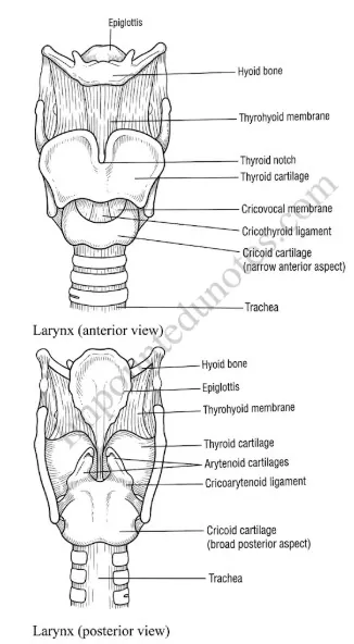
Figure: Larynx (anterior and posterior view)
Structure of larynx
The larynx (voice box) is the hollow cartilaginous box at the top of the trachea, in the neck from the 3rd to the 6th cervical vertebra (C3-C6).
Cause of Adam’s apple
At puberty, testosterone enlarges the thyroid cartilage in males. It projects at the front of the neck as the prominent Adam’s apple.
Meaning
Pulmonary ventilation (breathing) is inhaling oxygen from the external environment into the lungs and throwing out carbon dioxide in the reverse direction. An adult breathes 12-16 times per minute, by the action of the diaphragm and intercostal muscles.
Mechanism
| Inspiration | Expiration |
|---|---|
| Intrapulmonary pressure becomes less than atmospheric pressure. | Intrapulmonary pressure becomes more than atmospheric pressure. |
| External intercostal muscles contract; ribs move upward and outward. | Internal intercostal muscles contract; ribs move downward and inward. |
| Diaphragm contracts and becomes flat and straight. | Diaphragm relaxes and returns to dome shape. |
| Thoracic cavity and lungs expand; air rushes in. | Thoracic cavity and lungs decrease in size; air is pushed out. |
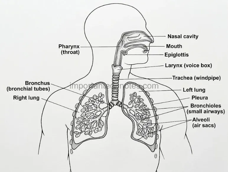
Figure: Human respiratory organs
The lungs are the primary respiratory organs.
Figure: Exchange of oxygen and carbon dioxide between alveolus and blood capillary by diffusion
Simple diffusion along the partial pressure gradient (from higher to lower partial pressure) is responsible. Alveoli are the primary sites of gaseous exchange.
Transport of O2
Transport of CO2
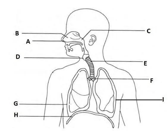
Figure: Human respiratory system with parts labelled A to I
i. Labels
| Label | Part | Label | Part |
|---|---|---|---|
| A | External nostril | F | Bronchus |
| B | Nasal cavity | G | Right lung |
| C | Pharynx | H | Diaphragm |
| D | Larynx (voice box) | I | Left lung |
| E | Trachea (wind pipe) |
ii. Function of D and H
iii. Difference between G and I
| G (right lung) | I (left lung) |
|---|---|
| Larger, with 3 lobes. | Slightly smaller, with 2 lobes. |
| Right bronchus is wider and about 2.5 cm long; divides into 3 branches. | Left bronchus is about 5 cm long; divides into 2 branches. |
| No cardiac notch. | Has a cardiac notch for the heart. |
Respiratory System Diagram: Long Answer Questions
The organs of respiration are the nose, pharynx, larynx, trachea, bronchi and bronchioles, lungs and the respiratory muscles.
Figure: Path of air in the human respiratory system from nostrils to alveoli
1. Nose (nostrils and nasal chambers)
The first organ, opening by external nostrils and to the pharynx by internal nostrils. A septum divides it into two chambers lined with ciliated epithelium and goblet cells. It has the vestibule, respiratory region (3 pairs of nasal conchae) and olfactory region. Four paranasal sinuses (frontal, ethmoidal, maxillary, sphenoid) open into it. Functions: filters, humidifies and warms air, detects smell and adds to voice resonance.
2. Pharynx
A short tube behind the buccal cavity, common to food and air, with nasopharynx, oropharynx and laryngopharynx. Internal nostrils and the eustachian tube open into the nasopharynx.
3. Larynx
The voice box with nine cartilages and vocal cords (see Short Q.1).
4. Trachea
About 12 cm long and 2.5 cm wide, from C6 to the 5th thoracic vertebra, ventral to the oesophagus. It is lined by ciliated pseudostratified epithelium with goblet cells and held open by 16-20 C-shaped cartilaginous rings.
5. Bronchi and bronchioles
The trachea divides at the 5th thoracic vertebra into a right bronchus (wider, 2.5 cm; 3 branches) and a left bronchus (5 cm; 2 branches). They divide into tertiary bronchi, bronchioles, terminal and respiratory bronchioles, alveolar ducts and alveoli.
6. Lungs
Paired, conical, spongy organs in pleural sacs, with about 300 million alveoli. Type I pneumocytes carry out gaseous exchange; type II make surfactant.
Figure: Human respiratory organs
7. Respiratory muscles
The diaphragm and external and internal intercostal muscles bring about breathing. Neck, shoulder and abdominal muscles are accessory muscles.
Respiration includes the taking in of O2, its transport, the oxidation of food in tissues and the removal of CO2.
Figure: Four steps of respiration: pulmonary ventilation, gaseous exchange, transport of gases and internal respiration
1. Pulmonary ventilation
Breathing in (inspiration, intrapulmonary pressure lower than atmosphere: external intercostals contract, diaphragm flattens, thorax expands) and breathing out (expiration, pressure higher: internal intercostals contract, diaphragm returns to dome shape). 12-16 breaths per minute.
2. Gaseous exchange
In alveoli, by simple diffusion. PO2 100 mmHg (alveoli) → 40 mmHg (venous blood); PCO2 44 mmHg (blood) → 40 mmHg (alveoli).
3. Transport of gases
O2: 1-3% dissolved in plasma and 97-99% as oxyhaemoglobin. CO2: about 7% as carbonic acid, 70% as bicarbonates (chloride shift) and 23% as carbamino-haemoglobin.
4. Internal respiration
Near the cells, oxyhaemoglobin dissociates (high PCO2 and temperature, low pH): HbO2 → Hb + O2. O2 diffuses into cells, where food is oxidised to release energy: C6H12O6 + 6O2 → 6CO2 + 6H2O + energy (38 ATP in aerobic respiration). CO2 made returns to the lungs.
4. Respiratory System Diagram PDF
PDF Notes 1: Human Biology (Unit 8)
PDF Notes 2: Respiratory System (Chapter 2)
5. Respiratory System Diagram: Frequently Asked Questions
What are the main parts of the respiratory system?
Where can I see a respiratory system diagram?
What are the respiratory system parts and functions?
What are the 5 functions of the respiratory system?
What are the types of respiration?
What is the difference between inspiration and expiration?
How are oxygen and carbon dioxide transported in blood?
Where can I download the respiratory system PDF for Class 12 Biology?
Is this Class 12 Biology book PDF for Nepal NEB?
Students also search for: