
Development of Frog Notes
Class 12 Biology Notes • NEB Syllabus 2083
Development of frog notes for Class 12 Biology Unit 7 Developmental Biology: syllabus, micro syllabus, 11 solved multiple choice questions, all short and long answers with diagrams on spermatogenesis, oogenesis, cleavage, blastula, gastrulation, neural tube and coelom, and three PDF notes for the NEB syllabus.
1. Development of Frog Notes Syllabus
Unit 7: Developmental Biology
This page covers chapter 7.2 of Unit 7 Developmental Biology of the NEB Class 12 Biology syllabus.
Micro syllabus: 7. Developmental Biology
Read more about the frog, gametogenesis, cleavage and gastrulation on Wikipedia. More notes: Class 12 Biology notes, Class 12 Chemistry notes and Class 12 Physics notes.
2. Development of Frog Notes: Multiple Choice Questions
Question 1. What is the final product of spermatogenesis?
- a) A viable sperm
- b) Four sperms
- c) Four viable eggs
- d) A sperm and three polar bodies
Correct Answer: b) Four sperms
Reason: One primary spermatocyte completes meiosis I and II and gives four haploid spermatids, and each becomes a sperm. The polar bodies belong to oogenesis.
Question 2. In which body organ does gametogenesis occur?
- a) Liver
- b) Gonads
- c) Kidney
- d) Heart
Correct Answer: b) Gonads
Reason: Gametes are formed in the gonads: sperms in the testes and ova in the ovaries.
Question 3. What is the main function of the acrosome in the sperm of frog?
- a) To provide energy for movement
- b) Storing genetic material
- c) Nourishing sperms
- d) To penetrate the egg
Correct Answer: d) To penetrate the egg
Reason: The acrosome on the head holds sperm lysin (hyaluronidase), which dissolves the egg membrane. Energy comes from the mitochondria of the middle piece.
Question 4. Which part of the egg in frog contains yolk?
- a) Animal pole
- b) Perivitelline space
- c) Vegetal pole
- d) Albuminous layer
Correct Answer: c) Vegetal pole
Reason: The frog egg is telolecithal: yolk is packed toward the vegetal pole, while the animal pole has the nucleus and black pigment.
Question 5. Which of the following hormone stimulate ovulation in frog?
- a) Oestrogen
- b) Progesterone
- c) Testosterone
- d) Leutinizing hormone
Correct Answer: d) Leutinizing hormone
Reason: As the chapter states, the pituitary gonadotropin (LH) triggers the release of the mature egg from the ovary.
Question 6. Which of the following structure surrounds the developing oocytes of frog?
- a) Sertoli cells
- b) Chorion
- c) Follicular cells
- d) Corpus luteum
Correct Answer: c) Follicular cells
Reason: Follicular cells form the follicle around each growing oocyte. Sertoli cells are in the testis.
Question 7. What triggers the completion oogenesis in frog?
- a) Hormonal changes
- b) Male croaking sound
- c) Cleavage
- d) Entry of sperm into egg
Correct Answer: d) Entry of sperm into egg
Reason: The secondary oocyte is shed in meiosis II and completes it only after the sperm enters (second polar body forms).
Question 8. What does the formation of grey crescent signify in frog’s fertilization?
- a) Site of sperm entry
- b) Makes embryo asymmetrical
- c) Marks the position of dorsal lip of blastopore
- d) Involves the formation of blastocoel
Correct Answer: c) Marks the position of dorsal lip of blastopore
Reason: The grey crescent forms opposite the sperm entry point and marks where the dorsal lip of the blastopore will appear. It does turn the radial egg into a bilateral one, but the chapter states the blastopore point.
Question 9. Which of the following describes role of blastocoel in the blastula stage of development?
- a) It is the site where ectoderm forms.
- b) It is the cavity filled with yolk.
- c) The fluid filled cavity which provides space for cell differentiation and development.
- d) It is the place where neural tube forms.
Correct Answer: c) The fluid filled cavity which provides space for cell differentiation and development.
Reason: The blastocoel is filled with albuminous fluid, not yolk, and gives room for the cell movements of gastrulation.
Question 10. What is the major event of epiboly of frog gastrulation?
- a) Mesodermal cell moves inward to form archenteron
- b) Ectodermal cells spread to cover the entire surface of embyo
- c) Cell of endoderm involute to form yolk plug
- d) Dorsal lip of blastopore form.
Correct Answer: b) Ectodermal cells spread to cover the entire surface of embyo
Reason: In epiboly the pigmented micromeres divide and spread over the megameres of the vegetal pole, leaving only the yolk plug uncovered.
Question 11. When the embryo of frog is exposed to a chemical that inhibits signaling molecules involved in the formation of mesoderm. What effect would this likely have on coelom formation in the embryo?
- a) The embryo would form an ectodermal coelom instead of a mesodermal coelom.
- b) The mesoderm would fail to develop, and the embryo would lack a coelom.
- c) The coelom would form normally, but the mesoderm would remain undifferentiated.
- d) The embryo would form an expanded coelom but with defects in the organ systems.
Correct Answer: b) The mesoderm would fail to develop, and the embryo would lack a coelom.
Reason: The coelom is a schizocoel formed by splitting of the lateral plate mesoderm, so no mesoderm means no coelom.
3. Development of Frog Notes: Short and Long Answer Questions
Short Answer Questions
Oogenesis is the formation of the mature female gamete (ovum) in the ovary, under the control of hormones such as FSH and LH. It has three phases.
Figure: Oogenesis in frog
One primary oocyte finally gives one functional ovum and 2-3 polar bodies, which degenerate because they take no part in reproduction.
Identification: the figures are not printed with the question in the exercise file. They are taken as the two gametes of frog: A = sperm and B = ovum (egg).
| Feature | A (Sperm) | B (Ovum) |
|---|---|---|
| Size | Very small, microscopic | Large, 1.75-2 mm in diameter |
| Shape and parts | Elongated, with head, middle piece and tail | Spherical, with animal and vegetal hemispheres |
| Movement | Motile, swims with the tail | Non-motile |
| Yolk | No yolk | Yolk present (mesolecithal, telolecithal) |
| Covering | No jelly coat, acrosome on the head | Vitelline membrane and a jelly-like albumen layer |
| Number | Produced in millions | Produced in fewer numbers (but many in spawn) |
If your paper shows the unfertilized and the fertilized egg (the two figures at the end of chapter 7.1), the differences are:
| Feature | Unfertilized egg | Fertilized egg |
|---|---|---|
| Grey crescent | Absent | Present, opposite the sperm entry |
| Symmetry | Radially symmetrical | Bilaterally symmetrical |
| Fertilization membrane | Absent | Present, with a perivitelline space |
| Polar bodies | Absent (first polar body only) | Polar bodies visible, second maturation completed |
The stage is called the blastula. Because its cells are many-layered, the frog blastula is also called a coeloblastula.
Formation (blastulation):
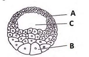
Figure: Vertical section of the late blastula (A micromeres, B macromeres, C blastocoel)
i. Blastula (late blastula, vertical section).
ii. A = micromeres (roof, animal hemisphere); B = megameres or macromeres (floor, vegetal hemisphere); C = blastocoel.
iii. Differences between A and B:
| A (Micromeres) | B (Megameres) |
|---|---|
| Small cells | Large cells |
| Pigmented, free of yolk | Unpigmented, full of yolk |
| Divide rapidly | Divide slowly |
Figure: Vertical section of the late blastula (A micromeres, B macromeres, C blastocoel)
Long Answer Questions
Spermatogenesis is the process of formation of haploid, motile sperms from diploid germ cells in the seminiferous tubules of the testis. It is controlled by FSH and testosterone and has three phases.
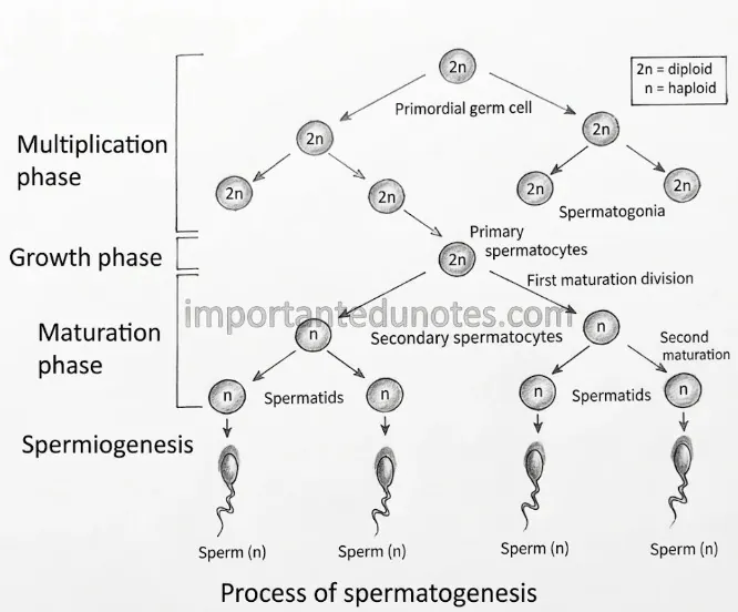
Figure: Chart of spermatogenesis
Spermiogenesis (spermateliosis): the spermatids change into sperms. A tail grows, the acrosome forms from the Golgi body, the nucleus becomes narrow and the cytoplasm is reduced. In frog, the sperms are released into the lumen of the seminiferous tubules in bundles; this release is called spermiation.
Structure of sperm: head with acrosome and a haploid nucleus, a middle piece with centrioles and mitochondria (the energy chamber), and a tail with 9+2 microtubules.
Cleavage is the repeated mitotic division of the zygote that forms a multicellular embryo without any increase in its size. It starts 2-3 hours after fertilization. In frog it is holoblastic (complete) and unequal, because the yolk lies toward the vegetal pole and slows division there.
| Cleavage | Plane | Result |
|---|---|---|
| 1st | Meridional (vertical), from animal to vegetal pole | 2 equal blastomeres |
| 2nd | Meridional, at right angle to the 1st | 4 equal blastomeres |
| 3rd | Horizontal (latitudinal), a little above the equator | 8 cells: 4 micromeres above, 4 megameres below |
| 4th | Meridional, two synchronous divisions | 16 cells |
| 5th | Horizontal, two synchronous divisions | 32 cells: 16 micromeres and 16 megameres |
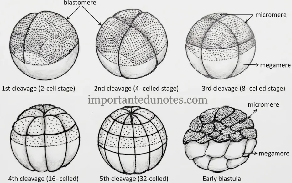
Figure: Cleavage in frog (2-cell, 4-cell, 8-cell, 16-cell, 32-cell stage and early blastula)
After the fifth cleavage the solid mulberry-like ball of cells is the morula (morulation). It has no blastocoel. A cavity then appears and the embryo becomes the blastula.
Gastrulation is the series of cell movements (morphogenetic movements) that changes the blastula into a gastrula with the three primary germ layers: ectoderm, mesoderm and endoderm. Prof. Vogt studied it in frog by vital staining.
Major events of gastrulation:
Result: outer micromeres become ectoderm, inner megameres become endoderm, and the chordamesoderm forms the mesoderm and notochord in between.
| Germ layer | Gives rise to |
|---|---|
| Ectoderm | Epidermis, brain and spinal cord, olfactory and auditory organs, lens, pituitary, enamel of teeth |
| Mesoderm | Notochord, muscles, bone and cartilage, blood and vessels, dermis, excretory and reproductive system |
| Endoderm | Lining of the alimentary canal, liver, pancreas, lungs, thyroid, urinary bladder lining |
The ectoderm forms the nervous system in amphibians. Neural tube formation is called neurulation.
Figure: Formation of neural tube (neurulation)
The notochord below the neural plate supports the embryo and induces the plate to form.
Coelom is the cavity between the body wall and the gut. In frog it forms by splitting of the mesoderm, so it is a true coelom and is also called schizocoel.
Formation of the coelom:
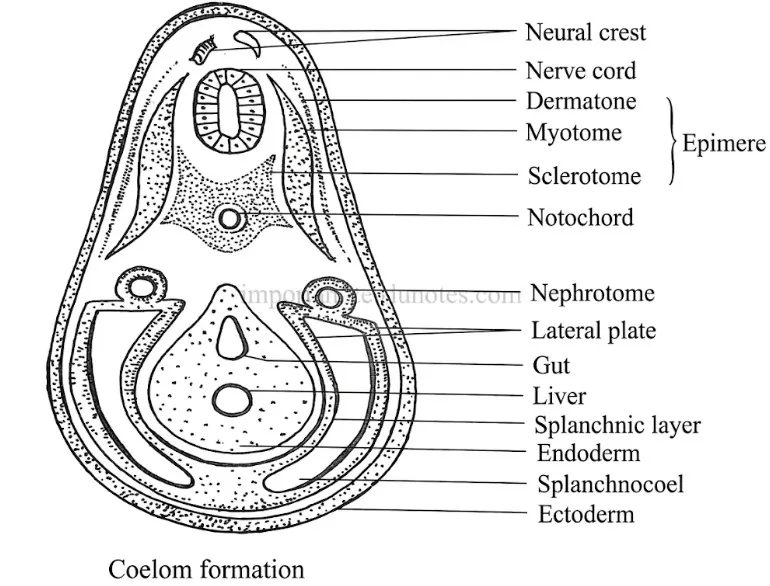
Figure: Formation of the coelom (schizocoel) in frog
The dorsal, thick mesoderm near the notochord is the epimere. It forms the mesodermal somites with a small cavity, the myocoel, which differentiates into the dermatome (dermis), myotome (muscles) and sclerotome (vertebral skeleton).
4. Development of Frog Notes PDF
PDF Notes 1: Developmental Biology (Unit 7)
PDF Notes 2: Gametogenesis (Chapter 7.1)
PDF Notes 3: Development of Frog (Chapter 7.2)
5. Development of Frog: Frequently Asked Questions
What are the Development of frog notes for Class 12 Biology? (Development of frog notes in simple words)
What are the stages of development of frog? (Development of frog stages)
Where can I get a development of frog pdf, development of frog diagram, development of frog ppt and development of frog slideshare?
What is the embryonic development of frog in Class 12 notes?
What is gametogenesis? (Gametogenesis notes, gametogenesis pdf)
What is gametogenesis in humans and gametogenesis in plants?
Where can I find a gametogenesis diagram, gametogenesis ppt and gametogenesis slideshare, and how is gametogenesis pronounced?
What is Developmental Biology? (Developmental biology notes, developmental biology syllabus)
Where can I get a developmental biology pdf, developmental biology book and developmental biology ppt?
What is the Developmental Biology journal and its Impact Factor?
Where can I get Class 12 Biology Book PDF Nepal and Class 12 Biology Book PDF Asmita Publication?
Where can I get Class 12 biology notes, Class 12 biology pdf and Class 12 Biology Notes NEB for this chapter?
Where can I get Class 12 Biology notes in the style of Note Mandir?
Students also search for: