
Digestive System Notes
Class 12 Biology Notes • NEB Syllabus 2083
Digestive system notes for Class 12 Biology Unit 8 Human Biology chapter 8.1: syllabus, micro syllabus, 12 solved multiple choice questions, all short and long answers with diagrams on salivary glands, tooth, liver, pancreas, small intestine, digestion and absorption, and two PDF notes for the NEB syllabus.
1. Digestive System Notes Syllabus
Unit 8: Human Biology
This page covers chapter 8.1 of Unit 8 Human Biology of the NEB Class 12 Biology syllabus.
Micro syllabus: 8. Human Biology
Read more about the human digestive system, digestion, salivary glands and the pancreas on Wikipedia. More notes: Class 12 Biology notes, Class 12 Chemistry notes and Class 12 Physics notes.
2. Digestive System Notes: Multiple Choice Questions
Question 1. Which of the following type of gland is present in stomach?
- a) Branched tubular
- b) Coiled tubular
- c) Simple tubular
- d) Alveolar
Correct Answer: a) Branched tubular
Reason: Gastric glands of the stomach mucosa are branched tubular glands that open into the stomach lumen.
Question 2. What is the hardest substance of human body?
- a) Enamel
- b) Ossein
- c) Dentine
- d) Chondrin
Correct Answer: a) Enamel
Reason: Enamel, the cover of the tooth crown, is the hardest substance of the body. Dentine is softer and forms the bulk of the tooth.
Question 3. Which part of the alimentary canal is chiefly responsible for the absorption of nutrients?
- a) Small intestine
- b) Stomach
- c) Large intestine
- d) Pancreas
Correct Answer: a) Small intestine
Reason: About 90% of absorption takes place in the small intestine, which has villi and microvilli. The pancreas is a gland, not part of the canal.
Question 4. Which of the following is the main function of oesophagus?
- a) Digestion of food particle
- b) Passage of swallowed food
- c) Absorption
- d) Mastication
Correct Answer: b) Passage of swallowed food
Reason: The oesophagus carries the bolus to the stomach by peristalsis. No digestion occurs in it.
Question 5. Which of the following enzyme changes trypsinogen into trypsin?
- a) Enterokinase
- b) Pepsin
- c) Sucrase
- d) Hydrochloric acid
Correct Answer: a) Enterokinase
Reason: Enterokinase from the intestinal mucosa activates trypsinogen. HCl activates pepsinogen in the stomach.
Question 6. Which of the following duct is also called duct of Santorini?
- a) Main pancreatic duct
- b) Accessory pancreatic duct
- c) Pancreatic ductule
- d) Hepatopancreatic duct
Correct Answer: b) Accessory pancreatic duct
Reason: The main pancreatic duct is the duct of Wirsung. The accessory duct is the duct of Santorini.
Question 7. Phagocytic cells in liver are called …………
- a) Kupffer cells
- b) Peyer’s patches
- c) Hepatocytes
- d) Ito cells
Correct Answer: a) Kupffer cells
Reason: Kupffer cells in the hepatic sinusoids engulf microbes, dead cells and foreign matter. Ito cells store fat and vitamin A.
Question 8. What is the duct of sub-mandibular salivary gland called?
- a) Wharton’s duct
- b) Duct of Rivinus
- c) Stenson’s duct
- d) Duct of Santorini
Correct Answer: a) Wharton’s duct
Reason: Wharton’s duct drains the submandibular gland. Stenson’s duct is of the parotid and the ducts of Rivinus are of the sublingual gland.
Question 9. Which of the following sentence best describes the impact of HCl in the stomach on enzymatic activity?
- a) HCl inhibit working of enzymes.
- b) HCl slow down the digestion of fats
- c) HCl activates pepsinogen to pepsin and aid in digestion of protein
- d) HCl reduces the effectiveness of enzymes.
Correct Answer: c) HCl activates pepsinogen to pepsin and aid in digestion of protein
Reason: HCl gives the acidic medium and converts inactive pepsinogen into active pepsin.
Question 10. A person with chronic diarrhea is losing a significant amount of electrolyte and water. Which part of the digestive system is mainly responsible for absorbing water?
- a) Small intestine
- b) Stomach
- c) Large intestine
- d) Oesophagus
Correct Answer: c) Large intestine
Reason: The textbook names the large intestine as the part mainly involved in absorbing water and salts from the undigested residue. Diarrhoea means this reabsorption fails.
Question 11. Where is bile produced in the body?
- a) Gall bladder
- b) Liver
- c) Lungs
- d) Hepatic duct
Correct Answer: b) Liver
Reason: Hepatic cells make the bile. The gall bladder only stores it.
Question 12. Which of the following is the characteristic of human teeth?
- a) Heterodont, acrodont and polyphyodont
- b) Heterodont, thecodont and polyphyodont
- c) Homodont acrodont and diphyodont
- d) Heterodont, thecodont and diphyodont
Correct Answer: d) Heterodont, thecodont and diphyodont
Reason: Human teeth are of different shapes (heterodont), fixed in sockets (thecodont) and come in two sets, milk and permanent (diphyodont).
3. Digestive System Notes: Short and Long Answer Questions
Short Answer Questions
The spacious cavity behind the mouth is the buccal cavity (oral cavity). The mouth first opens into a narrow space, the vestibule, between the lips and cheeks outside and the teeth and gums inside. The vestibule opens into the buccal cavity, which is lined by stratified squamous epithelium.
Structures present in the buccal cavity
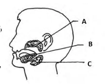
Figure: The three pairs of salivary glands
a) A = parotid gland (near the ear); B = sublingual gland (under the tongue); C = submandibular (sub-mandibular) gland (at the angle of the lower jaw).
b) All three secrete saliva, a slightly acidic (pH 6.8) fluid with water, salts, salivary amylase, lingual lipase, lysozyme, mucus and immunoglobulin.
c) Ducts:
| Gland | Duct | Share of saliva |
|---|---|---|
| A Parotid | Stenson’s duct (opens opposite the upper second molar) | 25% |
| B Sublingual | Ducts of Rivinus | 5% |
| C Submandibular | Wharton’s duct | 70% |
A tooth fixed in the jaw socket shows three parts in a longitudinal section: crown, neck and root.
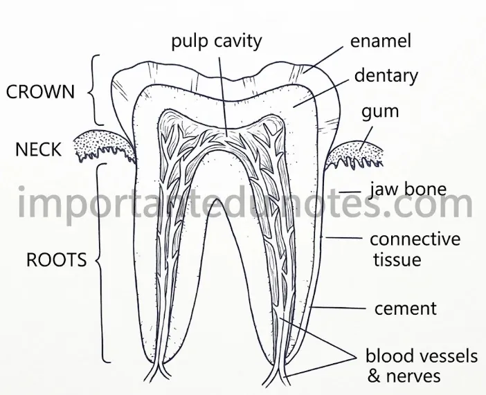
Figure: L.S. of a human tooth
The liver produces bile juice. It is the largest gland of the body, reddish-brown, weighs about 1.5 kg and lies on the right side of the abdomen just below the diaphragm.
Bile is alkaline (pH 8.6). It has no enzymes, but its bile salts emulsify fats, neutralize the acidic chyme and help absorb fats and fat-soluble vitamins.
In the duodenum the acidic chyme is mixed with bile, pancreatic juice and intestinal juice (succus entericus). This makes the medium alkaline, which suits the enzymes.
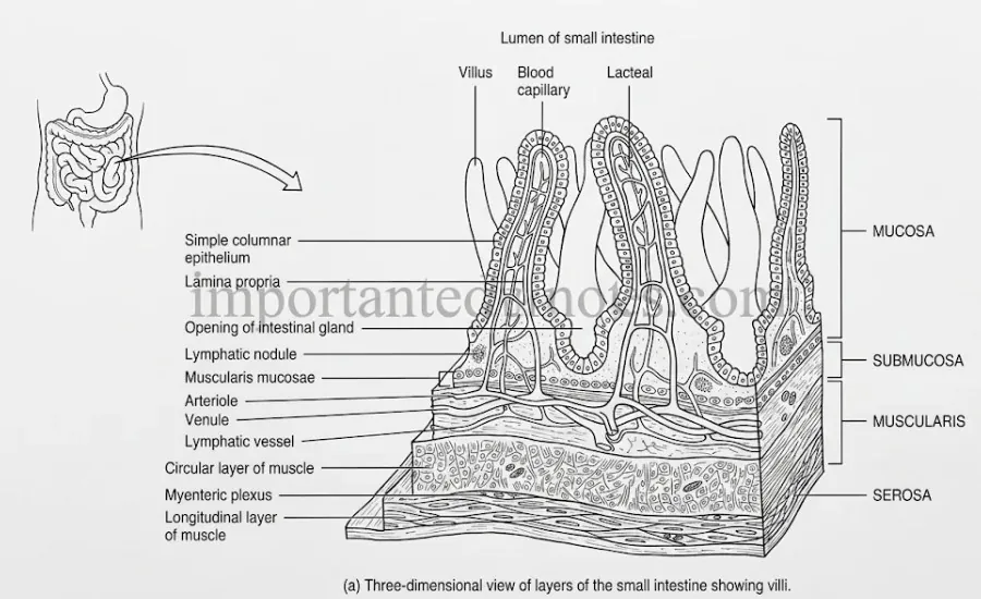
Figure: Wall of the small intestine with villi
Elastin → Dipeptides [elastase]
Starch → Maltose [pancreatic amylase]
Emulsified fats → Fatty acids + glycerol [pancreatic lipase]
Nucleic acids → Nucleotides [nucleases]
Intestinal juice completes the digestion at the brush border:
Maltose → Glucose + Glucose [maltase]
Lactose → Glucose + Galactose [lactase]
Sucrose → Glucose + Fructose [sucrase]
Fat → Fatty acids + glycerol [lipase]
Nucleotides → Nucleosides → Nitrogenous base + sugar [nucleotidase, nucleosidase]
Absorption is the process by which the end products of digestion pass through the mucosa of the alimentary canal into the blood or lymph. About 90% of it takes place in the small intestine through the villi, by simple diffusion, osmosis, facilitated diffusion and active transport.
| End product | How it is absorbed |
|---|---|
| Glucose, galactose (carbohydrate) | Active transport (with Na+), into the blood capillaries of the villi |
| Fructose | Facilitated diffusion through carrier proteins |
| Amino acids (protein) | Active transport into the blood capillaries; a small amount by simple diffusion |
| Fatty acids and glycerol (fat) | Simple diffusion. Fatty acids join bile salts and lecithin to form micelles, which enter the enterocytes. Inside, they are made into chylomicrons, which pass to the lacteals (lymph) and finally to the blood by the left subclavian vein |
Water is absorbed by osmosis, and vitamin B complex and C by simple diffusion. The large intestine absorbs some water, minerals and drugs.
Figure: Wall of the small intestine with villi
Protein → Peptones + proteoses [pepsin]
Casein → Paracasein [rennin, infants]
Paracasein + Ca++ → Calcium paracaseinate
Calcium paracaseinate → Peptones [pepsin]
Chymotrypsinogen → Chymotrypsin [trypsin]
Procarboxypeptidase → Carboxypeptidase [trypsin]
Proelastase → Elastase [trypsin]
Proteins, peptones, proteoses → Dipeptides [trypsin, chymotrypsin, carboxypeptidase]
Dipeptides → Amino acids [dipeptidase of succus entericus]
The amino acids are absorbed by the villi. No digestion occurs in the large intestine.
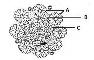
Figure: Section of pancreas (A acini, B connective tissue, C islet of Langerhans)
a) The figure is a section of the pancreas, a digestive gland. A = pancreatic acini (exocrine part, the flower-like clusters); B = connective tissue between the acini and the islet; C = islet of Langerhans (endocrine part).
b) Differences between A and C:
| A (Pancreatic acini) | C (Islet of Langerhans) |
|---|---|
| Exocrine part, forms about 99% of the pancreas | Endocrine part, forms the small remaining part |
| Has ducts (ductules) that carry the secretion | Ductless, hormones go directly into the blood capillaries |
| Cells are arranged around a central lumen | Cells (alpha, beta, delta, PP) form a compact mass |
| Secretes pancreatic juice | Secretes hormones |
c) Function of A: secretes the alkaline pancreatic juice (sodium bicarbonate and enzymes: trypsinogen, chymotrypsinogen, procarboxypeptidase, proelastase, amylase, lipase, nucleases) which digests carbohydrates, proteins, fats and nucleic acids in the duodenum. Function of C: alpha cells make glucagon, beta cells insulin, delta cells somatostatin and PP cells pancreatic polypeptide, which control the blood sugar and other functions.
Long Answer Questions
The digestive system is the group of organs that takes in food, digests it, absorbs it, assimilates it and egests the undigested part as faeces. It has the alimentary canal, the associated digestive glands and the physiology of digestion.
The alimentary canal of the digestive system is a long, coiled tube of 9-10 m, from mouth to anus.
| Organ | Structure | Function |
|---|---|---|
| Mouth and buccal cavity | Opening bounded by two lips; cavity with palate, tongue, teeth | Ingestion, chewing, tasting, mixing food with saliva |
| Pharynx | Vertical tube of 12 cm with nasopharynx, oropharynx and laryngopharynx; epiglottis guards the glottis | Common passage for food and air; swallowing |
| Oesophagus | Muscular tube of 25 cm, no serosa (tunica adventitia instead); cardiac sphincter at the lower end | Carries the bolus to the stomach by peristalsis; no digestion |
| Stomach | J-shaped bag, 30 cm × 15 cm; cardia, fundus, body and pylorus; gastric glands and rugae | Stores food 4-5 hours, churns it with gastric juice, starts protein digestion, forms chyme |
| Small intestine | About 6.5 m: duodenum (30 cm, C-shaped), jejunum (2.5 m), ileum (3.5 m); villi and microvilli | Main site of digestion and absorption |
| Large intestine | About 1.5 m: caecum with vermiform appendix, colon (ascending, transverse, descending, sigmoid) with haustra and taenia coli, rectum (15 cm) | Absorbs water and minerals; E. coli makes vitamin K and some B vitamins; stores faeces |
| Anal canal and anus | 2-4 cm canal with anal columns; internal (involuntary) and external (voluntary) sphincters | Egestion (defecation) of faeces |
The wall of the canal has four layers from outside to inside: serosa, muscularis (outer longitudinal and inner circular smooth muscle), submucosa and mucosa.
Figure: Wall of the small intestine with villi
Digestion is the conversion of complex food into simple, absorbable forms by hydrolytic enzymes. Carbohydrates, proteins and fats are changed into glucose, amino acids, and fatty acids with glycerol. Vitamins, minerals and water need no digestion.
1. Carbohydrates
Stomach: no carbohydrate digestion
Small intestine: Starch → Maltose [pancreatic amylase]
Maltose → Glucose + Glucose [maltase]
Lactose → Glucose + Galactose [lactase]
Sucrose → Glucose + Fructose [sucrase]
2. Proteins
Proteins → Peptones + proteoses [pepsin]
Small intestine: Trypsinogen → Trypsin [enterokinase]
Proteins, peptones, proteoses → Dipeptides [trypsin, chymotrypsin, carboxypeptidase]
Dipeptides → Amino acids [dipeptidase]
3. Fats
Bile salts emulsify fats in the duodenum
Emulsified fats → Fatty acids + glycerol [pancreatic lipase]
Fat → Fatty acids + glycerol [intestinal lipase]
Regulation: digestion is controlled by neural and hormonal mechanisms. The sight, smell and taste of food start the secretion of saliva and gastric juice through the nervous system. The gastrointestinal hormones are:
| Hormone | Source | Target organ | Action |
|---|---|---|---|
| Gastrin | Stomach | Stomach | Stimulates gastric glands to secrete gastric juice |
| Enterogastrone (GIP) | Duodenum | Stomach | Inhibits gastric secretion |
| Secretin | Duodenum | Pancreas, liver, stomach | Releases bicarbonate in pancreatic juice, increases bile secretion, decreases gastric secretion |
| Cholecystokinin | Small intestine | Gall bladder, pancreas | Contracts the gall bladder to release bile, stimulates pancreatic juice |
| Enterocrinin | Small intestine | Small intestine | Stimulates crypts of Lieberkuhn |
| Duocrinin | Duodenum | Duodenum | Stimulates Brunner’s glands |
4. Digestive System Notes PDF
PDF Notes 1: Human Biology (Unit 8)
PDF Notes 2: Digestive System (Chapter 8.1)
5. Digestive System: Frequently Asked Questions
What are the Digestive system notes for Class 12 Biology? (Digestive system in human)
What are the digestive system parts?
What is the digestive system function?
Where can I get a digestive system diagram and a digestive system drawing?
Can I get a digestive system paragraph for exams?
Is there a digestive system project and a digestive system for kids?
What is Human Biology? (Human biology notes, human biology anatomy)
Where can I get a Human Biology book, human biology course and human biology genetics notes?
Is there a Human biology 3D model, a Human biology app or Human Biology IGCSE notes?
Where can I get Class 12 Biology Book PDF Nepal and Class 12 Biology Book PDF Asmita Publication?
Where can I get Class 12 biology notes, Class 12 biology pdf and Class 12 Biology Notes NEB for this chapter?
Where can I get Class 12 Biology notes in the style of Note Mandir?
Students also search for: