
Circulatory System Notes
Class 12 Biology Notes • NEB Syllabus 2083
Circulatory system notes for Class 12 Biology Unit 8 Human Biology chapter 8.3: syllabus, micro syllabus, 20 solved multiple choice questions, all short and long answers with diagrams on the heart, heartbeat conduction, blood pressure, blood groups, arterial and venous systems, and two PDF notes for the NEB syllabus.
1. Circulatory System Notes Syllabus
Unit 8: Human Biology
This page covers chapter 8.3 of Unit 8 Human Biology of the NEB Class 12 Biology syllabus.
Micro syllabus: 8. Human Biology
Read more about the circulatory system, heart, cardiac cycle and blood groups on Wikipedia. More notes: Class 12 Biology notes, Class 12 Chemistry notes and Class 12 Physics notes.
2. Circulatory System Notes: Multiple Choice Questions
Question 1. Which of the following pericardium is closest to the heart?
- a) Parietal pericardium
- b) Endocardium
- c) Epicardium
- d) Fibrous pericardium
Correct Answer: c) Epicardium
Reason: The epicardium is the visceral pericardium, which covers the heart surface. The parietal layer is the outer, tough layer.
Question 2. Which of the following layer consist of cardiac muscles?
- a) Epicardium
- b) Endocardium
- c) Myocardium
- d) Pericardium
Correct Answer: c) Myocardium
Reason: The myocardium is the thick middle layer made of cardiac muscle fibres.
Question 3. What are the muscular ridges on the interior surface of atria called?
- a) Chordae corneae
- b) Musculi pectinati
- c) Papillary muscle
- d) Endocardium
Correct Answer: b) Musculi pectinati
Reason: Musculi pectinati are the pectinate muscle ridges of the atrial wall. Papillary muscles are in the ventricles.
Question 4. Tricuspid valve is situated in between…..
- a) Atria
- b) Right atrium and right ventricles
- c) Ventricles
- d) Left atrium and left ventricle
Correct Answer: b) Right atrium and right ventricles
Reason: The tricuspid valve guards the right atrio-ventricular opening. The left one has the bicuspid (mitral) valve.
Question 5. In embryo, Foramen ovale is the aperture lying in between right atrium and ………….
- a) Left atrium
- b) Aorta
- c) Right ventricle
- d) Pulmonary artery
Correct Answer: a) Left atrium
Reason: In the foetus the two atria communicate through the foramen ovale. After birth it closes and leaves the fossa ovalis.
Question 6. Which of the following statements describes the best about the role of SA node in the heart beat?
- a) It delays the electrical signal to allow complete atrial systole before ventricular systole
- b) It initiates electrical impulse for heart beat
- c) It is responsible for ventricular contraction only
- d) It checks the backflow of blood
Correct Answer: b) It initiates electrical impulse for heart beat
Reason: The SA node is the pacemaker. The AV node passes the impulse on, and valves check the backflow.
Question 7. Rashmi’s Grandfather is 65 years old who has been living with a dangerously slow heart beats. In this situation if he consults a cardiologist, he is possibly suggested to
- a) Do heavy physical exercise everyday
- b) Take less salty and spicy food
- c) Implant an artificial pacemaker
- d) Replace heart valves
Correct Answer: c) Implant an artificial pacemaker
Reason: A very slow beat means the SA node is not working well. An artificial pacemaker gives the impulses.
Question 8. Which of the following event is responsible for hearing lubb dupp sound during heart beat?
- a) Blood under pressure through aorta
- b) Closure of bicuspid and tricuspid valve
- c) Closing of semilunar valves
- d) Closure of bicuspid and tricuspid valves followed by semilunar valves
Correct Answer: d) Closure of bicuspid and tricuspid valves followed by semilunar valves
Reason: “Lubb” comes from the closing of the AV valves and “dupp” from the closing of the semilunar valves.
Question 9. To which of the following bundle of His transmits impulse of contraction?
- a) Atrium
- b) AV node
- c) SA node
- d) Purkinje fibre
Correct Answer: d) Purkinje fibre
Reason: The impulse goes from the AV node to the bundle of His and then to the Purkinje fibres of the ventricles.
Question 10. Which of the following statement is incorrect?
- a) Person having blood group A has antigen A in RBC and antibody b in plasma.
- b) Person having blood group B has antigen A in RBC and antibody a in plasma.
- c) Person having blood group AB has antigens A and B in RBC and no antibody in plasma.
- d) Person having blood group O has no antigen in RBC and antibodies a b in plasma.
Correct Answer: b) Person having blood group B has antigen A in RBC and antibody a in plasma.
Reason: Group B has antigen B (not A) on its RBCs and antibody a in plasma.
Question 11. John was unknown about his blood group, falls in an accident. He immediately needs blood transfusion. One of his friend having valid certificate of his own blood type offers blood without delay. What was the blood group of the donor?
- a) Blood group A
- b) Blood group AB
- c) Blood group B
- d) Blood group O
Correct Answer: d) Blood group O
Reason: Group O has no antigen on its RBCs, so it is the universal donor and can be given to any recipient.
Question 12. Through which blood vessel does the left auricle receive pure blood?
- a) Coronary vein
- b) Pulmonary artery
- c) Pulmonary vein
- d) Coronary artery
Correct Answer: c) Pulmonary vein
Reason: Two pairs of pulmonary veins bring oxygenated blood from the lungs to the left atrium.
Question 13. Which of the following valve is present at the opening of the coronary sinus?
- a) Mitral
- b) Tricuspid
- c) Eustachian
- d) Thebesian (coronary) valve
Correct Answer: d) Thebesian (coronary) valve
Reason: The opening of the coronary sinus is guarded by the Thebesian or coronary valve. The eustachian valve guards the inferior vena cava.
Question 14. From which of the following organ does the hepatic portal vein bring blood to the liver?
- a) Kidney
- b) Alimentary canal
- c) Heart
- d) Gonad
Correct Answer: b) Alimentary canal
Reason: The hepatic portal vein collects blood from the alimentary canal, pancreas and spleen.
Question 15. Which of the following is associated with decreased cardiac output?
- a) Parasympathetic signalling
- b) Hormones of adrenal medulla
- c) Pneumotaxic centre
- d) Sympathetic signalling
Correct Answer: a) Parasympathetic signalling
Reason: Parasympathetic fibres from the cardio-inhibitory centre slow the heart. Sympathetic fibres and adrenal hormones speed it up.
Question 16. Which of the following chemical is used to detect the presence of absence of Rh antigen in RBC of blood?
- a) Antiserum A
- b) Antiserum D
- c) Antiserum B
- d) Antiserum A and Antiserum B
Correct Answer: b) Antiserum D
Reason: Anti-D serum clumps Rh-positive RBCs. Anti-A and Anti-B test the ABO groups.
Question 17. Which of the following artery supplies oxygenated blood to diaphragm?
- a) Phrenic
- b) Pulmonary
- c) Carotid
- d) Vertebral
Correct Answer: a) Phrenic
Reason: The phrenic arteries (inferior phrenic from the descending aorta) supply the diaphragm.
Question 18. Which of the following vein is the longest vein in the human body?
- a) Renal
- b) Carotid
- c) Gonadal
- d) Saphenous
Correct Answer: d) Saphenous
Reason: The great saphenous vein of the leg is the longest vein. The carotid is an artery.
Question 19. Which phase of the cardiac cycle involves flow of blood from ventricles to different body parts?
- a) Atrial systole
- b) Isovolumetric contraction
- c) Ventricular systole
- d) Ventricular diastole
Correct Answer: c) Ventricular systole
Reason: In ventricular systole the ventricles contract and push blood into the aorta and the pulmonary artery.
Question 20. Mesenteric veins collect deoxygenated blood from
- a) Spleen
- b) Intestines
- c) Stomach
- d) Gonad
Correct Answer: b) Intestines
Reason: The mesenteric veins drain the intestines and pour into the hepatic portal vein.
3. Circulatory System Notes: Short and Long Answer Questions
Short Answer Questions
The human heart is a red, hollow, fibro-muscular, somewhat conical organ with a broad upper base and a narrow lower apex. It lies in the mediastinum of the thoracic cavity between the two lungs, slightly tilted to the left. It is about 12 cm long, weighs about 300 g and is the size of the owner’s clenched fist.

Figure: External structure of the human heart
Draw the longitudinal section like this and write the labels given below.

Figure: Internal structure of the human heart (L.S.)
Labels: superior vena cava, inferior vena cava, right atrium, left atrium, fossa ovalis, pectinate muscles, tricuspid valve, bicuspid (mitral) valve, chordae tendineae, papillary muscles, columnae carneae, right ventricle, left ventricle, interventricular septum, pulmonary trunk and pulmonary semilunar valve, aorta and aortic semilunar valve, pulmonary veins, epicardium, myocardium, endocardium.
The human heart is myogenic: the impulse for the heartbeat starts in the heart muscle itself (specialized nodal tissue), not in a nerve. A healthy heart beats about 72 times per minute.
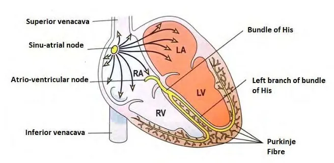
Figure: Origin and conduction of the heartbeat
Control: the cardiac centre in the medulla has the cardio-inhibitor (parasympathetic fibres, slows the heart) and the cardio-accelerator (sympathetic fibres, speeds it up). The adrenal medulla hormones (adrenaline, noradrenaline) and thyroxine also raise the heart rate.
Blood pressure is the pressure exerted by the blood on the wall of the blood vessel (mainly arteries) through which it flows. It is measured by a sphygmomanometer, with the brachial artery as reference. Normal value in an adult is about 120/80 mmHg: systolic 120 (ventricular systole) and diastolic 80 (ventricles relaxed). The difference, about 40 mmHg, is the pulse pressure.
| Hypotension | Hypertension |
|---|---|
| Persistent low blood pressure, as low as 90/50 mmHg | Persistent high blood pressure, beyond 140/90 mmHg |
| Caused by blood loss, vomiting, diarrhoea, heart problems, dilation of blood vessels | Caused by tension, fear, obesity, fatty food, smoking, nephritis |
| Poor blood supply, dizziness and weakness | Narrowing of the artery lumen; harms the brain, kidney and heart |
Causes of hypertension: tension and fear (stress), obesity, taking fatty food, smoking and nephritis (kidney disease), which lead to narrowing of the artery lumen.
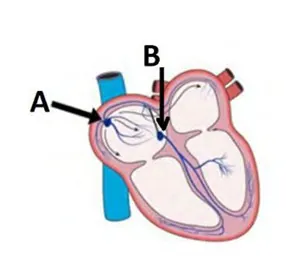
Figure: Conducting system of the heart (A SA node, B AV node)
i. A = sino-atrial (SA) node, near the opening of the superior vena cava; B = atrio-ventricular (AV) node.
ii. SA node: the natural pacemaker. It starts the impulse of the heartbeat (about 72 beats per minute) and makes the atria contract. AV node: picks up the impulse from the atria and passes it through the bundle of His and Purkinje fibres to the ventricles, so that they contract.
iii. If the SA node fails or works badly, an artificial pacemaker is implanted. It is a small battery-powered pulse generator placed under the collar bone, with leads and electrodes that give impulses to the heart muscle. Dr. Chardack first implanted it in 1960.
The arterial system is the system of arteries and their branches that carry pure blood from the heart to the body parts. The pulmonary artery is the exception and carries impure blood to the lungs.
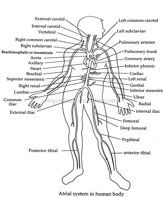
Figure: Arterial system in the human body
Blood grouping is the classification of blood by the presence or absence of specific antigens (agglutinogens) on the RBCs and antibodies (agglutinins) in the plasma. Karl Landsteiner (Nobel prize 1931) discovered the A, B and O groups, and the AB group was reported later by de Castello and Steini. The common systems are the ABO and the Rh systems.
| Group | Antigen on RBC | Antibody in plasma | Can donate to |
|---|---|---|---|
| A | A | b | A, AB |
| B | B | a | B, AB |
| AB | A and B | None (universal recipient) | AB |
| O | None | a and b | All groups (universal donor) |
Inheritance: three alleles (IA, IB, i) control the ABO group. Any person has two of them, so there are 6 genotypes and 4 phenotypes (A: IAIA or IAi; B: IBIB or IBi; AB: IAIB; O: ii).
Rh system: the Rh antigen (Rh factor) was found by Landsteiner and Weiner in 1940 in the Rhesus monkey. About 85% of people have it (Rh-positive), and the others are Rh-negative. Mixing Rh-positive blood with an Rh-negative person makes anti-Rh antibodies, causing agglutination. The first transfusion is a little dangerous and the second one may be fatal.
Detection: Anti-A, Anti-B and Anti-D antisera are used. Clumping (agglutination) shows that the antigen is present. Before transfusion the groups must be matched to avoid clumping, lysis of RBCs and circulatory failure.
Draw the venous system as given below, with the heart in the centre, and write the labels.
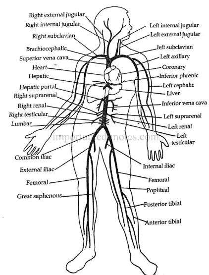
Figure: Venous system in the human body
Labels: right and left external jugular, internal jugular, subclavian, brachiocephalic, superior vena cava, heart, hepatic vein, hepatic portal vein, liver, suprarenal, renal, testicular (gonadal), lumbar, inferior vena cava, common iliac, external iliac, internal iliac, femoral, popliteal, great saphenous, anterior and posterior tibial veins.
Long Answer Questions
A myogenic heart is a heart in which the impulse for the beat starts in the cardiac muscle itself (the nodal tissue), not in the nerves. The human heart is myogenic.
Structure: the heart is four-chambered, enclosed in a pericardium, and its wall has epicardium, myocardium and endocardium.
Figure: Internal structure of the human heart (L.S.)
Working: double circulation. Blood passes through the heart twice in one complete round.
Pulmonary: Right ventricle → pulmonary arteries → lungs → pulmonary veins → left atrium
This keeps pure and impure blood from mixing, so the body gets blood rich in oxygen.
Origin and conduction of the heartbeat: SA node (pacemaker) → atria contract → AV node → bundle of His → Purkinje fibres → ventricles contract.
Cardiac cycle (about 0.8 s):
| Phase | Duration | Event |
|---|---|---|
| Atrial systole | 0.1 s | Both atria contract and push blood into the ventricles |
| Ventricular systole | 0.3 s | Ventricles contract and send blood to the lungs and the body. AV valves close (first sound, “lubb”) |
| Combined diastole | 0.4 s | All chambers relax and blood returns to the heart. Semilunar valves close (second sound, “dupp”) |
Cardiac output = heart rate × stroke volume = 72 × 70 ml = 5040 ml per minute.
The venous system is made of the veins and their branches that collect deoxygenated blood (except the pulmonary veins) from the body and bring it to the heart.
Figure: Venous system in the human body
Portal system in human: the hepatic portal system (a double capillary system). The hepatic portal vein collects blood from the alimentary canal, pancreas and spleen. It is formed by the cystic vein (gall bladder), splenic vein (spleen, pancreas, part of stomach), gastric vein (stomach and lower oesophagus), duodenal veins and mesenteric veins (intestines).
It is called a portal system because this nutrient-rich blood goes to the liver and not straight to the heart. Significance: the absorbed nutrients are processed, stored and used; toxins are detoxified; germs are destroyed by Kupffer cells; and ammonia from protein metabolism is converted to urea, which the kidney removes. (The renal portal system is found in the frog, not in human.)
4. Circulatory System Notes PDF
PDF Notes 1: Human Biology (Unit 8)
PDF Notes 2: Circulatory System (Chapter 8.3)
5. Circulatory System: Frequently Asked Questions
What are the Circulatory system notes for Class 12 Biology? (Circulatory system blood)
What are the circulatory system parts and circulatory system organs?
What is the function of the circulatory system?
Where can I get a circulatory system diagram and a circulatory system drawing?
Is there a circulatory system pdf?
What is the circulatory system for kids in simple words?
What is double circulation?
What is Human Biology? (Human biology notes, human biology anatomy)
Where can I get a Human Biology book, human biology course and human biology genetics notes?
Is there a Human biology 3D model, a Human biology app or Human Biology IGCSE notes?
Where can I get Class 12 Biology Book PDF Nepal and Class 12 Biology Book PDF Asmita Publication?
Where can I get Class 12 biology notes, Class 12 biology pdf and Class 12 Biology Notes NEB for this chapter?
Where can I get Class 12 Biology notes in the style of Note Mandir?
Students also search for: