
Excretory System Notes
Class 12 Biology Notes • NEB Syllabus 2083
Excretory system notes for Class 12 Biology Unit 8 Human Biology chapter 8.4: syllabus, micro syllabus, 12 solved multiple choice questions, all short and long answers with diagrams of the kidney, nephron and urine formation, and two PDF notes for the NEB syllabus.
1. Excretory System Notes Syllabus
Unit 8: Human Biology
This page covers chapter 8.4 of Unit 8 Human Biology of the NEB Class 12 Biology syllabus.
Micro syllabus: 8. Human Biology
Read more about the excretory system, kidney, nephron and urine on Wikipedia. More notes: Class 12 Biology notes, Class 12 Chemistry notes and Class 12 Physics notes.
2. Excretory System Notes: Multiple Choice Questions
Question 1. Which of the following nitrogenous compound is most toxic?
- a) Ammonia
- b) Urea
- c) Amino acid
- d) Uric acid
Correct Answer: a) Ammonia
Reason: Ammonia is highly toxic and needs a large volume of water to be removed. Uric acid is the least toxic.
Question 2. In the given animal, which one excrete amino acid without deamination?
- a) Earthworm
- b) Starfish
- c) Human
- d) Pigeon
Correct Answer: b) Starfish
Reason: Some echinoderms like starfish (and molluscs like Unio) excrete amino acids directly. This is aminotelism.
Question 3. Frogs’ tadpole is …….
- a) Ureotelic
- b) Ammonotelic
- c) Uricotelic
- d) Aminotelic
Correct Answer: b) Ammonotelic
Reason: The tadpole lives in water and excretes ammonia. The adult frog is ureotelic.
Question 4. What are terrestrial insects?
- a) Ureotelic
- b) Uricotelic
- c) Aminotelic
- d) Ammonotelic
Correct Answer: b) Uricotelic
Reason: Insects living on land save water by excreting uric acid as solid crystals.
Question 5. Which of the following is the position of human kidney?
- a) Intraperitoneal
- b) Extraperitoneal
- c) Retroperitoneal
- d) Interperitoneal
Correct Answer: c) Retroperitoneal
Reason: The kidneys lie behind the peritoneal cavity, so they are retroperitoneal.
Question 6. Which is not correct with respect to human kidney?
- a) Outer region is cortex and inner is medulla
- b) Bowman’s capsule confined to cortex
- c) Blood enters glomerulus through afferent venules
- d) The concave part of kidney is hilum
Correct Answer: c) Blood enters glomerulus through afferent venules
Reason: Blood enters the glomerulus through the afferent arteriole, not a venule.
Question 7. In which part, does the duct of Bellini open into?
- a) Hilum
- b) Renal pelvis
- c) Ureter
- d) Urinary bladder
Correct Answer: b) Renal pelvis
Reason: Ducts of Bellini run through the renal papillae and open into the renal pelvis.
Question 8. Which of the following part of nephron is responsible for obligatory reabsorption of water?
- a) Proximal convoluted tubule
- b) Distal convoluted tubule
- c) Loop of Henle
- d) Glomerulus
Correct Answer: a) Proximal convoluted tubule
Reason: About 80% of the water is absorbed in the PCT by osmosis, whether the person drinks much or little water.
Question 9. What is the cause of yellowish colour of urine?
- a) Urochrome
- b) Indole
- c) Skatole
- d) Biliverdin
Correct Answer: a) Urochrome
Reason: Urochrome (urobilin) pigment gives the yellow colour. Indole and skatole are faecal compounds.
Question 10. Which of the following substance is most likely to be absorbed from glomerular filtrate during the process of urine formation?
- a) Urea
- b) Glucose
- c) Sodium
- d) Potassium
Correct Answer: b) Glucose
Reason: Glucose is actively and fully reabsorbed in the PCT. Urea is only partly absorbed, and K+ is secreted.
Question 11. Which of the following is a characteristic unique to juxtamedullary nephrons compared to cortical nephrons?
- a) They have shorter loop of Henle that stay within cortex
- b) They are more abundant in the kidney than cortical nephron
- c) They do not participate in the formation of concentrated urine
- d) They have long loop of Henle that extend deep into the medulla
Correct Answer: d) They have long loop of Henle that extend deep into the medulla
Reason: Juxtamedullary nephrons have a long loop of Henle with vasa recta, which helps to make concentrated urine.
Question 12. The muscular layer of urinary bladder responsible for the contraction during micturition is known as
- a) Mucosa
- b) Detrusor muscle
- c) Tunica adventitia
- d) Urethral sphincter
Correct Answer: b) Detrusor muscle
Reason: The detrusor is the well-developed muscular layer that contracts to expel urine.
3. Excretory System Notes: Short and Long Answer Questions
Short Answer Questions
Excretion is the removal of metabolic wastes (nitrogenous compounds, excess water, salts, CO2, hormones) from the body. It keeps the body fluids and tissues in balance (homeostasis).
| Type | Waste | Examples |
|---|---|---|
| Ammonotelic | Ammonia: very toxic, very soluble, needs much water | Protozoans, sponges, Hydra, tapeworm, leech, aquatic arthropods and molluscs, freshwater bony fishes, tadpole |
| Ureotelic | Urea: less toxic and less soluble, can stay in the body for some time | Man, other mammals, adult amphibians, elasmobranchs (shark), marine bony fish, turtles |
| Uricotelic | Uric acid: least toxic, insoluble, passed as solid crystals, saves water | Insects, land crustaceans (woodlouse), land gastropods, snakes, lizards, birds |
| Aminotelic | Amino acids that the body cannot use are passed out as they are | Unio, Limnaea, Asterias (starfish) |
Some animals, such as the earthworm, lung fishes, African toad and crocodiles, show dual excretion: they pass both ammonia and urea.
| Ammonotelic | Ureotelic | Uricotelic | Aminotelic |
|---|---|---|---|
| Rahu | Shark | Pigeon | Lemnae (Limnaea) |
| Crustacean (aquatic) | Bat | Lizard | Starfish |
| Common carp | Cow | Snake | Unio |
| Tadpole | Human | Dragonfly | |
| Crab | Frog | Sparrow | |
| Horse | Woodlouse |
Dual excretion (ammonia and urea): earthworm and crocodile. The textbook lists both under dual excretion, so they are not put in one of the four columns. Lizard is written twice in the question and is counted once.
Draw the longitudinal section of the kidney as given below, and write the labels.
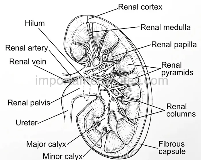
Figure: L.S. of the human kidney
Labels: renal capsule (fibrous capsule), renal cortex, renal medulla, renal pyramids, renal columns (of Bertini), renal papilla, minor calyx, major calyx, renal pelvis, hilum, renal artery, renal vein, ureter.
External structure: kidneys are paired, dark red, bean-shaped organs on either side of the vertebral column at the level of T12 to L3. They are retroperitoneal. The right kidney lies a little lower because of the liver. Each is about 11 cm long, 5.5 cm wide and 3 cm thick, and weighs about 150 g in male and 135 g in female.
Figure: L.S. of the human kidney
Internal structure (L.S.): there are two regions. The outer, darker cortex (about 1/3 of the tissue) has the Malpighian bodies, PCT and DCT. The inner, lighter and striated medulla (about 2/3) forms 15-16 conical renal pyramids, with the loop of Henle and the collecting ducts. The cortex goes between the pyramids as renal columns (of Bertini). The pyramids open by their papillae into the minor calyces, which join into major calyces, and then into the funnel-like renal pelvis, which leads into the ureter. Millions of nephrons are the functional units.
Ultrafiltration (glomerular filtration) is the purely physical filtration of blood plasma under pressure from the glomerulus into the Bowman’s capsule. It was first described by Richard Bright in the 19th century. It occurs in the Malpighian body (glomerulus and Bowman’s capsule) in the renal cortex.
Net filtration pressure (NFP):
NFP = 55 − (30 + 15) = 10 mmHg (or 7.3 − (4 + 2) = 1.3 kPa)
The rate of filtrate formation by both kidneys is the glomerular filtration rate (GFR): about 125 ml per minute, or about 180 litres a day.
Tubular secretion is the active process in which unwanted solutes and excess K+ are secreted from the peritubular capillaries (blood) into the tubular fluid. It helps to control the blood pH. It occurs in the PCT, DCT and collecting duct.
Animals benefitted: marine fishes and desert amphibians, which lack the glomerulus and Bowman’s capsule (aglomerular kidney). They cannot filter the blood, so they depend on tubular secretion to remove wastes.
Urine is the liquid waste formed by the kidneys.
Micturition (urination) is the discharge of urine from the bladder through the urethra. It is a reflex. The bladder fills by the sympathetic nerves. When it has about 300 ml of urine, the stretch receptors in its wall send impulses to the spinal cord and the micturition centre in the pons. Parasympathetic motor neurons then make the detrusor muscle contract and the internal sphincter relax. The external sphincter also relaxes (voluntary control) and the urine goes out.
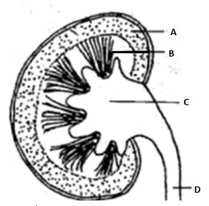
Figure: L.S. of kidney (A renal cortex, B renal medulla, C renal pelvis, D ureter)
i. A = renal cortex; B = renal medulla (renal pyramid); C = renal pelvis; D = ureter.
ii. Differences between A and B:
| A (Cortex) | B (Medulla) |
|---|---|
| Outer region, darker and granular | Inner region, lighter, with a striated look |
| About 1/3 of the kidney tissue | About 2/3 of the kidney tissue |
| Has Bowman’s capsule, PCT and DCT | Has loop of Henle, collecting ducts and ducts of Bellini |
| Continues inward between the pyramids as renal columns | Forms 15-16 conical renal pyramids |
iii. C (renal pelvis): the funnel-shaped space that receives urine from the major calyces and passes it to the ureter. D (ureter): a narrow tube of 25-30 cm that carries urine from the kidney to the urinary bladder by peristalsis. A fold of mucous membrane at its opening works as a valve and stops the backflow.
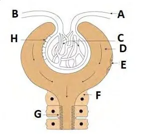
Figure: Malpighian body (renal corpuscle) with the start of the proximal tubule
a. The figure is the Malpighian body (renal corpuscle) joining the proximal tubule. A = efferent arteriole; B = afferent arteriole; C = glomerulus (capillary tuft); D = parietal layer of Bowman’s capsule; E = squamous epithelial cell of Bowman’s capsule; F = cell of the proximal convoluted tubule; G = microvilli (brush border); H = podocytes (visceral layer).
b. Differences between A and B:
| A (Efferent arteriole) | B (Afferent arteriole) |
|---|---|
| Narrower in diameter | Wider in diameter |
| Takes blood out of the glomerulus, to the peritubular capillaries | Brings blood into the Bowman’s capsule to form the glomerulus |
c. Glomerulus: the tuft of capillaries where the blood is filtered under pressure. Its wall has pores (fenestrae), so water and small solutes pass into the Bowman’s capsule.
d. Microvilli (brush border): they increase the surface area of the PCT cells for selective reabsorption. These cells also have many mitochondria for active transport.
Long Answer Questions
The major excretory organs of human are a pair of kidneys, a pair of ureters, a urinary bladder and a urethra.
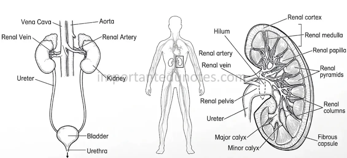
Figure: Excretory organs in the human body
Other organs that help in excretion: the skin (sweat and sebum), the lungs (CO2 and water vapour), the liver (urea is formed here; bile pigments go out with faeces) and the intestine (undigested matter, excess iron, calcium, indole and skatole).
The structural and functional unit of the kidney is the nephron (uriniferous tubule). Each kidney has about one million nephrons. Urine is formed in three steps.
1. Ultrafiltration in the Malpighian body: because the afferent arteriole is wider than the efferent arteriole, the blood pressure is high (glomerular hydrostatic pressure 55 mmHg). It is opposed by the blood osmotic pressure (30 mmHg) and the capsular pressure (15 mmHg), so NFP = 55 − (30 + 15) = 10 mmHg. Blood plasma without cells and proteins enters the Bowman’s capsule as the glomerular filtrate (GFR 125 ml per minute, about 180 L a day).
2. Selective reabsorption (useful substances go back to the blood):
| Part | What happens |
|---|---|
| PCT | Main site, about 70% of the filtrate is absorbed. About 65% of Na+ is actively absorbed. Glucose, amino acids, vitamins, lactic acid and HCO3− are absorbed actively. About 80% of the water follows by osmosis (obligatory reabsorption). K+, Cl−, Ca++, Mg++ and about 50% of urea are absorbed by diffusion. |
| Loop of Henle | The descending limb is highly permeable to water and not to salts. Water leaves, and the filtrate becomes hypertonic. The ascending limb is impermeable to water but actively transports Na+, K+ and Cl− out, so the filtrate becomes hypotonic and the medulla stays concentrated. |
| DCT | Facultative reabsorption: Na+ by aldosterone, Ca++ by parathormone and water by ADH. The filtrate is isotonic to plasma. |
| Collecting duct | More water is absorbed under ADH, Na+ under aldosterone, and some urea, Cl− and HCO3− too. |
3. Tubular secretion: creatinine, drugs, ammonia, H+, K+ and NH4+ are actively secreted from the blood into the PCT and DCT. Urea also enters the thin segment of the loop of Henle by diffusion.
The concentrated urine then flows through the collecting duct to the duct of Bellini, the renal pelvis, the ureter and the bladder.
A nephron is a long, thin and much coiled tubule, about 3-5.5 cm long. It has two parts: the Malpighian body and the renal tubule.
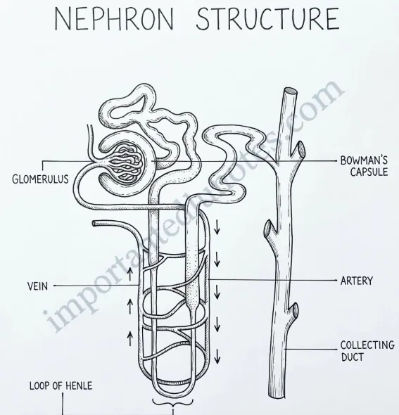
Figure: Structure of a nephron (schematic)
| Cortical nephron | Juxtamedullary nephron |
|---|---|
| About 80-85% of the nephrons | About 15-20% of the nephrons |
| Smaller and confined to the cortex | Larger, in both the cortex and the medulla |
| Short loop of Henle, not deep into the medulla | Long loop of Henle, deep into the medulla |
| No vasa recta | Has vasa recta (peritubular capillaries along the loop) |
| Controls the plasma volume at normal water intake | Controls the plasma volume at low water intake |
4. Excretory System Notes PDF
PDF Notes 1: Human Biology (Unit 8)
PDF Notes 2: Excretory System (Chapter 8.4)
5. Excretory System: Frequently Asked Questions
What are the Excretory system notes for Class 12 Biology? (Excretory system pdf)
What are the excretory system parts and excretory system organs?
What is the human excretory system and what does the kidney do?
Can you give an excretory system example?
What is the excretory system pronunciation?
What is Human Biology? (Human biology notes, human biology anatomy)
Where can I get a Human Biology book, human biology course and human biology genetics notes?
Is there a Human biology 3D model, a Human biology app or Human Biology IGCSE notes?
Where can I get Class 12 Biology Book PDF Nepal and Class 12 Biology Book PDF Asmita Publication?
Where can I get Class 12 biology notes, Class 12 biology pdf and Class 12 Biology Notes NEB for this chapter?
Where can I get Class 12 Biology notes in the style of Note Mandir?
Students also search for: