
Endocrinology Notes Class 12
Class 12 Biology Notes • NEB Syllabus 2083
Endocrinology notes for Class 12 Biology Unit 8 chapter 8.7: syllabus, micro syllabus, 16 solved multiple choice questions, short and long answers on endocrine glands, hormones and hormonal disorders, and two PDF notes.
1. Endocrinology Syllabus
Unit 8: Human Biology (this page: chapter 8.7 Endocrinology)
These Endocrinology notes follow the NEB Class 12 Biology syllabus (+2 Biology, Nepal), chapter 8.7 of Unit 8 Human Biology. Practise the multiple choice questions first, then work through the solved short and long answer questions, and finish with the PDF notes for quick revision.

Figure: Endocrinology notes, Class 12 Biology (NEB)
Micro syllabus: 8. Human Biology
Endocrinology is the study of the endocrine system. Read more about endocrinology, the endocrine system, hormones and the pituitary gland on Wikipedia. More notes: Class 12 Biology notes, Class 12 Chemistry notes and Class 12 Physics notes.
2. Endocrinology: Multiple Choice Questions
Question 1. Which of the following is an endocrine gland?
- a) Salivary gland
- b) Sweat gland
- c) Thyroid
- d) Tear gland
Correct Answer: c) Thyroid
Reason: The thyroid is ductless and releases hormones into blood. Salivary, sweat and tear glands are ducted exocrine glands.
Question 2. Who discovered the first hormone, secretin?
- a) William M. Bayliss and Ernest H. Starling
- b) Addison
- c) Cushing
- d) Starling
Correct Answer: a) William M. Bayliss and Ernest H. Starling
Reason: Secretin, secreted by intestinal mucosa to stimulate pancreatic juice, was found jointly by Bayliss and Starling.
Question 3. Which of the following is the common chemical nature of hormones and enzymes?
- a) Protein
- b) Steroid
- c) Carbohydrates
- d) Inorganic compound
Correct Answer: a) Protein
Reason: Enzymes are proteins, and many hormones (pituitary, pancreatic) are proteins too. Steroids are only one class of hormones.
Question 4. Where is the location of the pituitary gland?
- a) In the cerebellum
- b) Below the hypothalamus
- c) Above the hypothalamus
- d) In the medulla oblongata
Correct Answer: b) Below the hypothalamus
Reason: It lies in the sella turcica of the sphenoid bone, joined to the hypothalamus above it by the infundibulum.
Question 5. Which of the following hormones is secreted by the posterior lobe of the pituitary gland?
- a) Somatostatin
- b) Oxytocin
- c) Thyroid
- d) Prolactin
Correct Answer: b) Oxytocin
Reason: The posterior lobe stores and releases oxytocin and ADH. Prolactin comes from the anterior lobe and somatostatin from the hypothalamus.
Question 6. Which of the following is the function of the anti-diuretic hormone?
- a) Stimulates milk secretion
- b) Promotes the growth of muscle
- c) Regulates water balance
- d) Controls metabolism
Correct Answer: c) Regulates water balance
Reason: ADH increases water absorption in the DCT and collecting duct of the nephron.
Question 7. Which hormone stimulates the thyroid gland to secrete T3 and T4?
- a) TSH
- b) LH
- c) ACTH
- d) GH
Correct Answer: a) TSH
Reason: Thyroid stimulating hormone from the anterior pituitary acts on the thyroid. ACTH acts on the adrenal cortex.
Question 8. Which part of the pituitary gland releases the hormone synthesized by the neurosecretory cells of the hypothalamus?
- a) Anterior
- b) Posterior
- c) Middle
- d) Both anterior and middle
Correct Answer: b) Posterior
Reason: The posterior lobe makes no hormone. It only stores and releases oxytocin and vasopressin made in the hypothalamus.
Question 9. If a patient has a swollen thyroid gland, which test is commonly used to check whether the thyroid gland is over-secreting or under-secreting?
- a) CT scan
- b) Urine test
- c) Stool test
- d) Blood test for T3, T4, and TSH
Correct Answer: d) Blood test for T3, T4, and TSH
Reason: Blood levels of T3, T4 and TSH show whether the gland is over- or under-active.
Question 10. Which of the following functions is carried out by calcitonin?
- a) Increases the blood calcium level
- b) Decreases the blood calcium level
- c) Increases the blood sugar level
- d) Stimulates the production of erythrocytes
Correct Answer: b) Decreases the blood calcium level
Reason: Calcitonin lowers blood calcium by increasing calcium deposition in bones and reducing its reabsorption from urine.
Question 11. Which of the following disorders results from the over-secretion of the thyroid gland?
- a) Myxoedema
- b) Graves’ disease
- c) Cushing’s syndrome
- d) Acromegaly
Correct Answer: b) Graves’ disease
Reason: Graves’ disease (exophthalmic goitre) is hyperthyroidism. Myxoedema is hypothyroidism in adults.
Question 12. Which of the following hormones is not produced by the adrenal cortex?
- a) Cortisol
- b) Adrenaline
- c) Aldosterone
- d) Androgen
Correct Answer: b) Adrenaline
Reason: Adrenaline is made by the adrenal medulla. The cortex makes cortisol, aldosterone and androgens.
Question 13. What is the main function of cortisol?
- a) Regulating blood pressure
- b) Controlling metabolism and stress response
- c) Stimulating muscle growth
- d) Lowering the blood pressure
Correct Answer: b) Controlling metabolism and stress response
Reason: Cortisol, the stress hormone, regulates carbohydrate, protein and fat metabolism and mobilizes energy in stress.
Question 14. Which two minerals are balanced by aldosterone in the body?
- a) Calcium and phosphorus
- b) Sodium and magnesium
- c) Sodium and potassium
- d) Calcium and potassium
Correct Answer: c) Sodium and potassium
Reason: Aldosterone increases Na⁺ and water absorption and K⁺ excretion in the kidneys.
Question 15. Addison’s disease is caused by the deficiency of which hormone?
- a) Adrenaline
- b) Aldosterone
- c) Cortisol
- d) Aldosterone and cortisol
Correct Answer: d) Aldosterone and cortisol
Reason: It results from deficiency of both mineralocorticoids (aldosterone) and glucocorticoids (cortisol) of the adrenal cortex.
Question 16. Which of the following conditions causes Graves’ disease?
- a) Hyposecretion of adrenal gland
- b) Hypersecretion of thyroid gland
- c) Hyposecretion of thyroid gland
- d) Hypersecretion of adrenal gland
Correct Answer: b) Hypersecretion of thyroid gland
Reason: Excess T3 and T4 give a high metabolic rate, weight loss and protruding eyeballs.
3. Endocrinology: Short and Long Answer Questions
Endocrinology: Short Answer Questions
| Exocrine glands | Endocrine glands |
|---|---|
| Ducted glands; secretion flows through a duct. | Ductless glands; secretion goes directly into the blood stream. |
| Secretions are called enzymes. | Secretions are called hormones. |
| Site of action is near the site of secretion. | Site of action is far away from the site of secretion. |
| Examples: salivary gland, liver, sweat gland. | Examples: pituitary gland, thyroid gland. |
A gland that works as both is a heterocrine gland, for example the pancreas and gonads.
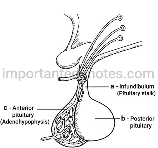
Figure: Pituitary gland and hypothalamus
Posterior lobe (neurohypophysis, pars nervosa) is the storage and releaser center.
Structure: It forms about 25% of the pituitary and develops from a downward growth of brain tissue. Axons of hypothalamic neurosecretory cells end here as axon terminals embedded in neuroglial cells called pituicytes. The hormones are stored in vesicles called Herring bodies.
Function: It secretes no hormone itself. It stores and releases oxytocin (uterine contraction in childbirth, milk ejection) and vasopressin or ADH (water absorption in the kidney, raises blood pressure).
| Hypothyroidism | Hyperthyroidism | |
|---|---|---|
| Cause | Less secretion of T3 and T4: iodine deficiency in diet (simple goitre), or autoimmune damage of the gland (Hashimoto’s disease). | Excess secretion of T3 and T4 (thyrotoxicosis), as in Graves’ disease. |
| Symptoms | Cretinism in children: dwarfism, mental deficiency, slow heartbeat, low metabolic rate. Myxoedema in adults: puffy face and hands, dry pale skin, dull look, low BP, low BMR and temperature. | Protruding eyeballs (exophthalmic goitre), insomnia, weight loss, restlessness, very high metabolic rate, fast heartbeat, palpitation and high temperature. |
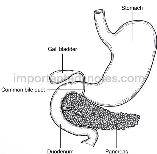
Figure: Pancreas: exocrine acini and endocrine islets of Langerhans
The pancreas is a soft, yellowish gland (about 15 cm, 60 g) just behind the stomach, in the curve of the duodenum. It is endodermal in origin.
Structure: About 99% is the exocrine part of lobules or acini with a central ductule. About 1% is the endocrine part, the islets of Langerhans (1–2 million cells), with four cell types: beta cells (about 60%), alpha cells (25%), delta cells (10%) and F or PP cells.
Function: Acini secrete pancreatic juice with digestive enzymes. Beta cells secrete insulin, which lowers blood sugar by converting glucose into glycogen and fat. Alpha cells secrete glucagon, which raises blood sugar by glycogenolysis. Delta cells secrete somatostatin, which regulates insulin and glucagon. F cells secrete pancreatic polypeptide.
Disorder: Low insulin causes diabetes mellitus (hyperglycemia and glycosuria).
Function: Four small glands on the back of the thyroid secrete parathormone (PTH) from their chief cells. PTH regulates calcium and phosphate levels between blood and other tissues.
Calcium balance: When blood calcium falls, PTH raises it to normal by drawing calcium from bones into plasma, increasing calcium absorption in the alimentary canal and reducing calcium loss in urine. Calcitonin of the thyroid does the opposite when calcium is high. Normal calcium is needed for muscle contraction, blood clotting and nerve impulses.
Disorders: Low PTH causes tetany; excess PTH causes hypercalcemia and kidney stones.
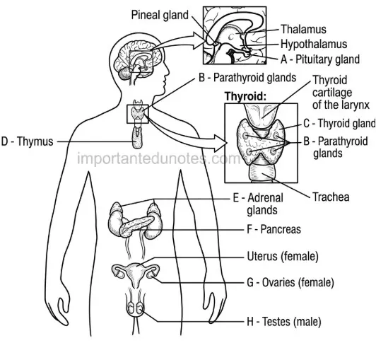
Figure: Human endocrine glands: ovary (G) and testis (H)
Location: The testes lie in the scrotum and the ovaries lie in the pelvic cavity on either side of the uterus. They are the primary reproductive organs and also heterocrine glands.
Ovary: Oestrogens bring growth of uterus and vagina, breast development, wider pelvis and female characters. Progesterone prepares the uterus for conception and stimulates mammary gland development in pregnancy.
Testis: Testosterone, the main androgen, develops male sex organs and secondary sexual characters (muscle and bone mass, body hair, broad shoulders, deep voice).
| Hormones | Enzymes |
|---|---|
| Secreted by endocrine glands. | Secreted by exocrine glands. |
| Secreted at one site and carried by blood to another site. | Site of action is generally near the site of secretion. |
| Low molecular weight. | Very high molecular weight. |
| May be steroids, proteins, peptides or amino acid derivatives. | Simple proteins. |
| Used up in their action. | Not used up in their action. |
| Act slowly or quickly. | Act slowly. |
| May slow down or accelerate reactions. | Speed up reactions. |
Structure: The adrenal medulla is the inner part of the adrenal gland, developed from embryonic ectoderm. It consists of rounded groups of large granular chromaffin cells. It is controlled directly by the sympathetic nervous system, not by the pituitary.
Function: It secretes adrenaline (epinephrine, 80%) and noradrenaline (norepinephrine, 20%), together called catecholamines. They help in the 3F response (fright, fight, flight). Adrenaline is a vasoconstrictor, raises cardiac output and blood pressure, causes glycogenolysis and breaks down fat. Noradrenaline mainly acts on blood vessels.
It is the pineal gland, a small pine-cone shaped gland (0.1–0.2 g) near the center of the brain between the two cerebral hemispheres.
It secretes melatonin, the sleep hormone, which regulates the sleep and wake cycle (circadian rhythm). Secretion increases in dim light and decreases in bright light. It possibly also inhibits growth of sex organs before puberty.
Figure: Diagram e: endocrine glands of the human body
e. i. Label the parts A, B, C, D, E, F, G, H.
| Part | Name |
|---|---|
| A | Pituitary gland |
| B | Thyroid gland |
| C | Parathyroid gland |
| D | Thymus gland |
| E | Adrenal gland |
| F | Pancreas |
| G | Ovary |
| H | Testis |
ii. Which of these part is also called gland of emergency?
Part E, the adrenal gland. Its medulla secretes adrenaline (emergency hormone) in fright, fight and flight.
iii. What is the function of D?
D is the thymus. It secretes thymosin, which promotes the proliferation and maturation of T-lymphocytes.
iv. What are the two differences between B and C?
| B (thyroid) | C (parathyroid) |
|---|---|
| Largest endocrine gland, butterfly shaped with two lobes joined by an isthmus. | Four small rounded bodies embedded on the back of the thyroid. |
| Secretes T3, T4 and calcitonin (lowers blood calcium). | Secretes parathormone (raises blood calcium). |
Figure: Diagram f: pituitary gland with hypothalamic neurons
f. i. Identify the gland? Write its specific location in our body.
It is the pituitary gland (hypophysis). It lies just below the hypothalamus in the sella turcica (hypophyseal fossa) of the sphenoid bone and is joined to the brain by the infundibulum.
ii. Label the parts a, b and c.
a = infundibulum (stalk carrying axons from the hypothalamus), b = posterior lobe (neurohypophysis), c = anterior lobe (adenohypophysis) with its capillary network.
iii. Write two secretions from part b and part c each.
Part b (posterior lobe): oxytocin and ADH (vasopressin), stored and released here.
Part c (anterior lobe): any two of TSH, ACTH, FSH, LH, prolactin, growth hormone.
Endocrinology: Long Answer Questions
The hypothalamus is the control centre or supreme commander of endocrine regulation, because it controls the pituitary gland.
a) Structure
It is the basal, almond-shaped region of the diencephalon of the forebrain, developed from embryonic ectoderm. It contains clusters of neurons called nuclei that secrete releasing and inhibiting hormones. It has three regions:
| Region | Main nuclei | Role |
|---|---|---|
| Anterior (supra-optic) | Supra-optic, paraventricular | Secretes CRH, TRH, GnRH, oxytocin, vasopressin, somatostatin; regulates body temperature (sweating) and circadian rhythm. |
| Middle (tuberal) | Ventromedial, arcuate | Ventromedial controls appetite; arcuate releases GHRH. |
| Posterior (mammillary) | Posterior hypothalamic, mammillary | Regulates temperature (shivering, stops sweat); mammillary nuclei relate to memory. |
b) Function of its secretions
| Hormone | Function |
|---|---|
| CRH | Makes pituitary release ACTH. |
| TRH | Makes anterior pituitary release TSH. |
| GnRH | Makes anterior pituitary release FSH and LH. |
| GHRH | Stimulates growth hormone secretion. |
| Somatostatin | Stops release of growth hormone and TSH. |
| Oxytocin | Childbirth and lactation. |
| Vasopressin (ADH) | Regulates water level of the body. |
Releasing hormones reach the anterior pituitary through the hypophyseal portal veins. Oxytocin and ADH pass through axons to the posterior lobe for storage.
The pituitary is called the master gland because its hormones regulate the functions of other endocrine glands (thyroid, adrenal cortex, gonads). Its own activity is controlled by the hypothalamus.
Figure: Pituitary gland as the master gland
Figure: Pituitary gland (hypophysis)
a) Structure
It is the smallest endocrine gland: oval, reddish brown, about 1.3 cm across and 500 mg, in the sella turcica of the sphenoid bone and attached to the hypothalamus by the infundibulum. It has dual origin: the adenohypophysis from Rathke’s pouch (foregut roof) and the neurohypophysis from brain tissue. It has three lobes: anterior (pars distalis), middle (pars intermedia) and posterior (pars nervosa). The anterior and middle lobes form the adenohypophysis (about 75%), with acidophils, basophils and chromophobes. The posterior lobe (about 25%) has axon terminals and pituicytes.
b) Functions of hormones
| Hormone | Source | Function |
|---|---|---|
| TSH | Anterior | Stimulates thyroid to secrete T3 and T4. |
| ACTH | Anterior | Stimulates adrenal cortex to secrete corticoids. |
| FSH | Anterior | Female: follicle maturation and oestrogen. Male: sperm production. |
| LH | Anterior | Female: ovulation and corpus luteum. Male: testosterone from Leydig cells. |
| Prolactin | Anterior | Growth of mammary glands and milk production. |
| Growth hormone | Anterior | Growth of bones and muscles; affects carbohydrate, protein and fat metabolism. |
| MSH | Middle | Darkens skin pigmentation (more in lower vertebrates). |
| Oxytocin | Posterior | Uterine contraction in labour; milk ejection. |
| ADH | Posterior | Water absorption in DCT and collecting duct; raises BP. |
c) Hormonal disorders
| Hormone | Hyposecretion | Hypersecretion |
|---|---|---|
| Growth hormone | Dwarfism (stunted growth, normal intelligence); Simmond’s disease | Gigantism (in childhood); acromegaly (in adults: large hands, feet and lower jaw) |
| ADH | Diabetes insipidus: large volume of dilute urine | Anti-diuresis: small volume of urine |
| Prolactin | – | Galactorrhoea, infertility, amenorrhoea (female); gynecomastia and sterility (male) |
The thyroid gland controls the basal metabolic rate (BMR) through T3 and T4.
a) Structure
It is the largest endocrine gland, butterfly or ‘H’ shaped, in the neck from C5 to T1 just below the larynx, dark red and highly vascular, endodermal in origin. It has two lobes on either side of the trachea joined by a connective tissue band, the isthmus. It is 3–7 cm long and weighs 20–25 g in a fibrous capsule. It contains thousands of thyroid follicles lined by cuboidal follicular cells and filled with iodinated colloid, thyroglobulin, which contains T3 and T4. Between the follicles lie parafollicular (C) cells that secrete calcitonin.
b) Hormones and functions
T3 and T4 (thyroxine) are made from iodine and tyrosine, under control of TSH. They maintain BMR and heat production (thermogenesis), regulate carbohydrate, protein and fat metabolism, are needed for tissue growth in childhood, regulate urine output through renal tubules, and are essential for metamorphosis in amphibians.
Calcitonin lowers blood calcium by increasing its deposition in bones and decreasing its reabsorption from urine.
c) Hormonal disorders
| Disorder | Cause | Features |
|---|---|---|
| Cretinism | Hypothyroidism in children | Dwarfism, mental deficiency, undeveloped sex organs, slow heartbeat, low BMR |
| Myxoedema | Hypothyroidism in adults | Puffy face and hands, dry pale skin, dull look, low BP, heartbeat, BMR and temperature |
| Simple goitre | Iodine deficiency | Swollen gland in the neck |
| Hashimoto’s disease | Autoimmune damage | Gland progressively damaged; less thyroxine |
| Graves’ disease | Hyperthyroidism | Protruding eyeballs, insomnia, weight loss, high BMR, heartbeat and temperature |
Location: The two adrenal (supra-renal) glands are small, triangular, yellowish glands, each lying on top of a kidney. Each is about 5 cm long and 5 g in weight, covered by a thin fibrous capsule.
Emergency gland: Adrenaline secretion rises greatly in anger, fright, heat or cold. Adrenaline and noradrenaline prepare the body for fright, fight and flight, so the gland (and adrenaline) is called the emergency gland.
a) Structure
Figure: Structure of the adrenal gland (L.S.)
The cortex (outer, about 80%, from mesoderm, rich in fatty and cholesterol cells) has three layers; the medulla (inner, from ectoderm) has chromaffin cells. The cortex is stimulated by ACTH.
b) Functions
| Hormone | Function |
|---|---|
| Aldosterone (mineralocorticoid) | Na⁺ and water absorption, K⁺ excretion in kidneys. |
| Cortisol (glucocorticoid) | Metabolism of carbohydrate, protein, fat; gluconeogenesis; anti-inflammatory; stress hormone. |
| Sex corticoids | Small amounts of androgens and oestrogen; sex organs and secondary characters. |
| Adrenaline | Vasoconstriction, higher cardiac output and BP, glycogenolysis, fat breakdown. |
| Noradrenaline | Mainly regulates blood vessels and circulation. |
c) Hormonal disorders
| Disorder | Cause | Features |
|---|---|---|
| Addison’s disease | Deficiency of mineralocorticoids and glucocorticoids | Low Na⁺, high K⁺, nausea, bronze pigmentation, low BP, weakness |
| Cushing’s disease | Excess cortisol | Abdominal obesity, moon face, poor wound healing, high blood sugar |
| Conn’s syndrome | Hypersecretion of aldosterone | High Na⁺, low K⁺, high BP, polyuria |
| Hypo-aldosteronism | Low aldosterone | Low Na⁺, dehydration, low BP |
| Adrenal virilism | Excess androgen in females | Beard, moustache, male voice |
| Gynaecomastia | Excess oestrogen in males | Enlarged breasts, female voice |
4. Endocrinology PDF Notes
PDF Notes 1: Human Biology (Unit 8)
PDF Notes 2: Endocrinology (Chapter 7)
5. Endocrinology: Frequently Asked Questions
What is Endocrinology? (Endocrinology notes Class 12)
How do pronunciation and meaning of Endocrinology go together?
What is the difference between exocrine and endocrine glands?
Why is the pituitary called the master gland?
Which tests are used in Endocrinology to check the thyroid?
What are common Endocrinology symptoms of thyroid disorders?
What is Endocrinology treatment, and where is Endocrinology near me?
Which glands secrete which hormones?
Where can I download Endocrinology PDF for Class 12 Biology?
Is this Class 12 Biology book PDF for Nepal NEB?
Students also search for: