
Sense Organs
Class 12 Biology Notes • NEB Syllabus 2083
Sense organs for Class 12 Biology Unit 8 Human Biology: syllabus, micro syllabus, 22 solved multiple choice questions, short and long answer questions on the human eye and ear, with figures and two PDF notes.
1. Sense Organs Syllabus
Unit 8: Human Biology (Chapter 8.6)
Sense organs are specialised organs with receptors that detect stimuli and send impulses to the CNS. The two most important are the eye and the ear. These notes follow the NEB Class 12 Biology syllabus (+2 Biology, Nepal), Unit 8, chapter 6. Practise the multiple choice questions, then the solved short and long answers with figures, then the PDF notes.

Figure: Sense organs, Class 12 Biology (NEB)
Micro syllabus: 8. Human Biology
Read more about sense organs, the human eye and the ear on Wikipedia. More notes: Class 12 Biology notes, Class 12 Chemistry notes and Class 12 Physics notes.
2. Multiple Choice Questions
Question 1. Photoreceptor of the vertebrate is
- a) eye
- b) ear
- c) nose
- d) tongue
Correct Answer: a) eye
Reason: The eye has rods and cones that detect light. Ear detects sound and balance; nose and tongue are chemoreceptors.
Question 2. An eye is extero-receptor, teloreceptor, special receptor, and
- a) photoreceptor
- b) chemoreceptor
- c) stato-acoustic receptor
- d) thigmo-receptor
Correct Answer: a) photoreceptor
Reason: The eye is sensitive to light. Stato-acoustic receptors belong to the ear and thigmo-receptors to the skin.
Question 3. Human eye is
- a) simple, monocular, and stereoscopic
- b) compound, binocular, and stereoscopic
- c) compound, monocular, and stereoscopic
- d) simple, binocular, and stereoscopic
Correct Answer: d) simple, binocular, and stereoscopic
Reason: Human eyes are simple (one lens each), both focus on the same object (binocular) and give 3-D (stereoscopic) vision.
Question 4. Nictitating membrane in man is
- a) upper eyelid
- b) lower eyelid
- c) vestigial eyelid
- d) well-developed eyelid
Correct Answer: c) vestigial eyelid
Reason: The third eyelid (nictitating membrane or plica semilunaris) is only a swelling in the inner corner of the eye.
Question 5. How many extraocular muscles are there in an eye?
- a) Two
- b) Four
- c) Six
- d) Eight
Correct Answer: c) Six
Reason: Four rectus (superior, inferior, internal, external) and two oblique (superior, inferior) muscles move the eyeball.
Question 6. The aperture of the pupil of eye is controlled by
- a) iris
- b) suspensory ligament
- c) ciliary body
- d) aquous humour
Correct Answer: a) iris
Reason: The iris has circular (sphincter) and radial (dilator) muscles that change the pupil size. The ciliary body holds and shapes the lens.
Question 7. In human eye, if rod cells contain the pigment rhodopsin then what type of pigment is found in cone cells?
- a) Iodopsin
- b) Opsin
- c) Lysozyme
- d) Retinal
Correct Answer: a) Iodopsin
Reason: Rods have rhodopsin (visual purple); cones have iodopsin-type pigments for bright light and colour vision.
Question 8. The sensory part of human eye is
- a) cornea
- b) choroid
- c) lens
- d) retina
Correct Answer: d) retina
Reason: The retina (nervous tunic) holds the rods and cones. Cornea and lens only refract light.
Question 9. If a person sees an object, image is formed on retina. Which one of the following sequence describes the correct path for light entering the human eye?
- a) Cornea – Lens – Pupil – Retina
- b) Cornea – Iris – Pupil – Retina
- c) Cornea – Pupil – Lens – Retina
- d) Cornea – Iris – Lens – Retina
Correct Answer: c) Cornea – Pupil – Lens – Retina
Reason: Light is refracted at the cornea, passes the pupil, is focused by the lens and falls on the retina.
Question 10. Vitamin A deficiency causes night blindness in person. In a night blinded person, which of the following are not functioning in low intensity of light?
- a) Lens
- b) Rods
- c) Cones
- d) Both rods and cones
Correct Answer: b) Rods
Reason: Vitamin A is needed to make rhodopsin of rods, which work in dim light. Cones need bright light.
Question 11. If a person using biconcave lens to correct vision, does not use glasses. Where will be the image formed in such man?
- a) Little behind retina
- b) Little in front of retina
- c) On fovea centralis
- d) On blind spot
Correct Answer: b) Little in front of retina
Reason: A concave lens corrects myopia, in which rays focus in front of the retina.
Question 12. Person who can see near object but cannot see far object has a type of eye defects known as
- a) myopia
- b) hypermetropia
- c) presbyopia
- d) diplopia
Correct Answer: a) myopia
Reason: Myopia (short-sightedness) gives clear near vision but blurred distant vision.
Question 13. Eustachian tube connects
- a) external ear with pharynx
- b) middle ear with pharynx
- c) internal ear with pharynx
- d) middle ear with internal ear
Correct Answer: b) middle ear with pharynx
Reason: It joins the tympanic cavity to the naso-pharynx and equalizes air pressure on both sides of the eardrum.
Question 14. In the middle ear of man, there are three ear ossicles. Identify and choose which of the following ear ossicles are arranged in correct sequence?
- a) Malleus – Incus – Saccule
- b) Malleus – Incus – Stapes
- c) Malleus – Stapes – Incus
- d) Malleus – Incus – Cochlea
Correct Answer: b) Malleus – Incus – Stapes
Reason: From the eardrum inward: malleus (hammer), incus (anvil), stapes (stirrup). Saccule and cochlea are not ossicles.
Question 15. The auditory ossicles are situated in
- a) external auditory meatus
- b) tympanic cavity
- c) bony labyrinth
- d) membranous labyrinth
Correct Answer: b) tympanic cavity
Reason: The ossicles form a chain inside the air-filled tympanic cavity of the middle ear.
Question 16. Which one of the following is associated with mechanical balance?
- a) Cochlea
- b) Cristae
- c) Tympanum
- d) Organ of Corti
Correct Answer: b) Cristae
Reason: Cristae (in ampullae of semicircular canals) and maculae detect movement and position. Cochlea and organ of Corti are for hearing.
Question 17. The sensory auditory part of the human ear is/are
- a) utriculus
- b) sacculus
- c) semicircular canals
- d) cochlea
Correct Answer: d) cochlea
Reason: The cochlea is the main organ of hearing. Utriculus, sacculus and canals are for balance.
Question 18. The organ of hearing is
- a) cristae
- b) semicircular canals
- c) organ of Corti
- d) helicotrema
Correct Answer: c) organ of Corti
Reason: The organ of Corti (spiral organ) has hair cells that start nerve impulses for hearing.
Question 19. In which part of the human body, is the organ of Corti present?
- a) Nasal chamber
- b) Retina
- c) Middle ear
- d) Internal ear
Correct Answer: d) Internal ear
Reason: It lies in the cochlea of the internal ear (labyrinth).
Question 20. The organ of Corti is situated in which part of the internal ear?
- a) Scala vestibule
- b) Scala media
- c) Scala tympani
- d) Macula and Cupula
Correct Answer: b) Scala media
Reason: It rests on the basilar membrane in the scala media (cochlear duct), which holds endolymph.
Question 21. Scala tympani is connected with scala vestibuli by
- a) Eustachian canal
- b) Scala media
- c) Helicotrema
- d) Fenestra ovalis
Correct Answer: c) Helicotrema
Reason: The two perilymph-filled canals meet at the apex of the cochlea through the helicotrema.
Question 22. The function of human ear is
- a) Vision
- b) Hearing
- c) Balance
- d) Both b and c
Correct Answer: d) Both b and c
Reason: The ear is a stato-acoustic organ: hearing (cochlea) and mechanical balance (vestibule and canals).
3. Short and Long Answer Questions
Sense Organs: Short Answer Questions
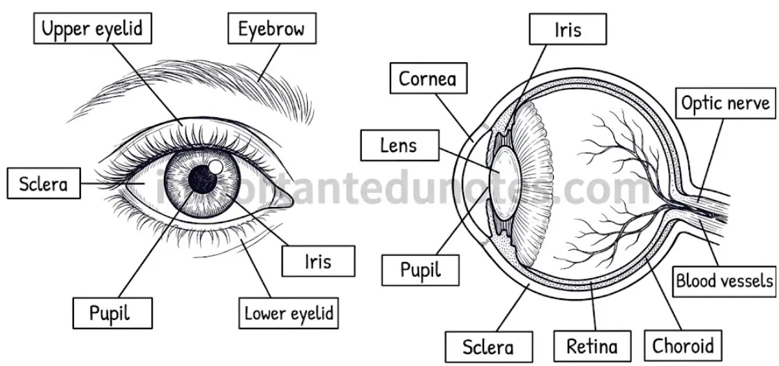
Figure: Internal and external structure of the human eye
Each eye is a hollow spherical eyeball (about 2.5 cm across) in a bony orbit. Its wall has three concentric tunics, and it holds the lens and two fluids.
Main structures
| Part | Structure and function |
|---|---|
| Fibrous tunic | Sclera (5/6, white, tough, protects and keeps shape) and transparent, avascular cornea (1/6, refracts light). |
| Vascular tunic (uvea) | Choroid (brown, absorbs light, supplies the retina), ciliary body (secretes aqueous humour, holds the lens) and iris (controls pupil size). |
| Nervous tunic (retina) | Light-sensitive; has rods and cones, the fovea centralis (cones only, sharpest vision) and the blind spot where the optic nerve leaves. |
| Lens | Transparent, biconvex, elastic; focuses light on the retina (accommodation). |
| Aqueous humour | Watery fluid between cornea and lens; nourishes and keeps the eye’s front shape. |
| Vitreous humour | Jelly-like fluid between lens and retina; keeps the back shape of the eye. |
Figure: Internal and external structure of the human eye
Functions
| Label | Function |
|---|---|
| Cornea | Transparent front part; refracts light into the eye. |
| Sclera | White outer coat; protects eyeball and keeps its shape. |
| Choroid | Pigmented, vascular; absorbs stray light and nourishes the retina. |
| Ciliary body | Secretes aqueous humour; ciliary muscles change the lens shape. |
| Iris and pupil | Iris controls the size of the pupil, which lets light pass. |
| Lens (with suspensory ligament) | Focuses light on the retina. |
| Retina | Rods (dim light) and cones (bright light, colour) start nerve impulses. |
| Fovea centralis | Centre of most distinct vision (cones only). |
| Blind spot | No rods or cones; optic nerve and blood vessels leave here. |
| Optic nerve | Carries impulses to the occipital lobe of the brain. |
Figure: Myopia and hypermetropia eye defects: image formed in front of and behind the retina
| Defect | Cause | Correction |
|---|---|---|
| Myopia (short-sight) | Eyeball long or lens too thick; image in front of retina; far objects blurred. | Concave lens |
| Hypermetropia (long-sight) | Eyeball short or lens loses elasticity; image behind retina; near objects blurred. | Convex lens |
| Astigmatism | Irregular cornea or lens; blurred vision at all distances. | Cylindrical lens |
| Presbyopia | Old age; lens loses flexibility. | Bifocal lens |
| Cataract | Clouding of the lens; first cause of blindness in Nepal. | Surgery, artificial lens |
| Glaucoma | Raised intraocular pressure damages the optic nerve; third cause of blindness in Nepal. | Medical treatment |
| Others | Corneal opacity (second cause of blindness in Nepal), macular degeneration, floaters, diabetic retinopathy, retinal detachment, dry eye, diplopia, colour blindness (more in men), amblyopia. | – |
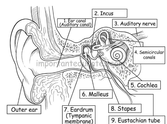
Figure: Structure of the human ear
| Part | Structure | Function |
|---|---|---|
| External ear | Pinna (funnel-shaped elastic cartilage), external auditory meatus (about 2.5 cm, S-shaped, with hairs and ceruminous glands) and tympanum (eardrum, 1 cm). | Collects sound, guides it to the eardrum, keeps dust out. |
| Middle ear | Air-filled tympanic cavity with three ossicles (malleus, incus, stapes), three muscles and three openings (Eustachian tube, oval window, round window). | Carries sound to the inner ear and raises its intensity 20 times; Eustachian tube equalizes pressure. |
| Internal ear | Bony labyrinth (perilymph) with the membranous labyrinth (endolymph): vestibule, semicircular canals and cochlea. | Balance (vestibule, canals) and hearing (cochlea). |
The membranous labyrinth floats in the perilymph of the bony labyrinth and is filled with endolymph. It is the true stato-acoustic organ with three parts:
Hearing
Pinna collects sound → meatus → eardrum vibrates → ossicles carry the vibrations (stapes fits the oval window) → perilymph → scala vestibuli → Reissner’s membrane → endolymph of scala media moves → tectorial membrane stimulates hair cells of the organ of Corti → impulses go by the auditory nerve to the temporal lobe.
Figure: Path of sound waves and nerve impulse in hearing through the human ear
Balance
When the body tilts or moves, endolymph and otoliths stimulate hair cells of maculae (static) and cristae (dynamic). Impulses reach the brain and the medulla oblongata detects the change; the brain then sends impulses to muscles to restore position.
Figure: Structure of the human ear
Functions
| Label | Function |
|---|---|
| Pinna (outer ear) | Collects sound waves and directs them inward. |
| External auditory meatus | Conducts sound to the eardrum; hairs and wax keep out dust. |
| Tympanic membrane | Vibrates with sound and starts the impulse. |
| Malleus, incus, stapes | Chain of ossicles; carry vibrations and increase intensity 20 times. |
| Eustachian tube | Joins middle ear to naso-pharynx; equalizes air pressure. |
| Semicircular canals (with vestibule) | Mechanical balance. |
| Cochlea | Hearing; has the organ of Corti. |
| Auditory nerve (CN VIII) | Carries impulses of hearing and balance to the brain. |
Long Answer Questions
The eye is the sense organ of vision. It is an extero-receptor, teloreceptor and special receptor with photoreceptors, one pair, simple, with binocular, trichromatic and stereoscopic (3-D) vision.
Figure: Internal and external structure of the human eye
Accessory structures
Main structures
Three tunics: fibrous (sclera and cornea; Schlemm’s canal drains aqueous humour), vascular (choroid, ciliary body, iris with pupil) and nervous (retina). The retina has ten layers. The rod and cone layer has rods with rhodopsin (dim light) and cones for red, green and blue colour. The fovea centralis has cones only; the blind spot has none. Contents: lens, aqueous humour and vitreous humour.
Function: how we see
Light passes the cornea, aqueous humour, pupil, lens and vitreous humour and is focused on the retina. The iris adjusts the pupil, and the lens changes its curvature (accommodation). Rods and cones form potential differences and an impulse, which the optic nerve carries to the visual cortex of the occipital lobe, where the image is recognised.
Figure: Path of light and nerve impulse in vision through the human eye
The ear is a stato-acoustic organ for mechanical balance and hearing. There is one pair, behind the cheek on each side of the head, connected to the brain by the auditory nerve (CN VIII).
Figure: Structure of the human ear
1. External ear
2. Middle ear
3. Internal ear (labyrinth)
Bony labyrinth filled with perilymph holds the membranous labyrinth filled with endolymph: vestibule (utriculus and sacculus with maculae), three semicircular canals (ampullae with cristae) and the cochlea (scala vestibuli, scala media with the organ of Corti, scala tympani, joined at the helicotrema). The organ of Corti has inner and outer hair cells, pillar cells and the overhanging tectorial membrane.
Functions
Balance: static (position of the head relative to gravity) and dynamic (sudden movements), by maculae and cristae, with the medulla oblongata sending corrective impulses. Hearing: sound is collected, the eardrum and ossicles carry it, fluids of the cochlea move, hair cells are stimulated and the auditory nerve carries impulses to the temporal lobe.
Figure: Path of sound waves and nerve impulse in hearing through the human ear
4. PDF Notes
PDF Notes 1: Human Biology (Unit 8)
PDF Notes 2: Sense Organs (Chapter 6)
5. Frequently Asked Questions
What are sense organs?
What are the five senses and their organs?
What is the function of the eye and ear?
Which part of the skin detects touch?
Where can I see drawings or diagrams of the eye and ear?
What are the types of receptors?
What is the difference between myopia and hypermetropia?
Which organ of the ear is the organ of hearing?
Where can I download the PDF for Class 12 Biology?
Is this Class 12 Biology book PDF for Nepal NEB?
Students also search for: