
Plant Anatomy Notes
Class 12 Biology Notes • NEB Syllabus 2083
Plant anatomy notes for Class 12 Biology Unit 1: syllabus, micro syllabus, 23 solved multiple choice questions, all short and long answer questions with labelled plant anatomy diagrams, two PDF notes and frequently asked questions.
1. Plant Anatomy Syllabus
Unit 1: Plant Anatomy Notes
These plant anatomy notes follow the NEB Class 12 Biology syllabus (+2 Biology, Nepal). Practise the multiple choice questions first, then work through the solved short and long answer questions with their diagrams, and finish with the PDF notes for quick revision.
Micro syllabus: 1. Plant Anatomy
Read more about plant anatomy, the meristem and secondary growth on Wikipedia. More notes: Class 12 Biology notes, Class 12 Chemistry notes and Class 12 Physics notes.
2. Plant Anatomy Notes: Multiple Choice Questions
Question 1. Plant anatomy deals with
- a) Heredity and variations
- b) Metabolic activities in plants
- c) Structure and function of plant tissues
- d) Reproduction in plants
Correct Answer: c) Structure and function of plant tissues
Reason: Plant anatomy is the study of the internal structure of plant parts, mainly their tissues. Metabolism belongs to plant physiology.
Question 2. Who is known as the ‘father of plant anatomy’?
- a) N. Grew
- b) G. Mendel
- c) R. Hooke
- d) Theophrastus
Correct Answer: a) N. Grew
Reason: Nehemiah Grew (1641-1712) made the first detailed microscopic study of plant parts and coined the terms tissue and parenchyma.
Question 3. Which tissue makes the outer lining of plant parts?
- a) Cambium
- b) Epidermis
- c) Pith
- d) Phloem
Correct Answer: b) Epidermis
Reason: Epidermis is the outermost protective layer of the young plant body. Cambium, pith and phloem lie inside.
Question 4. Meristematic cell has
- a) Large vacuole
- b) No nucleus
- c) No cell wall
- d) High rate of cell division
Correct Answer: d) High rate of cell division
Reason: Meristematic cells are young and keep dividing by mitosis. They have a big nucleus and dense cytoplasm, and vacuoles are absent or very small.
Question 5. Which is the correct pair?
- a) Intercalary meristem: root length
- b) Lateral meristem: plant height
- c) Apical meristem: inter-nodal length
- d) Root apex: root length
Correct Answer: d) Root apex: root length
Reason: The apical meristem at the root apex increases the length of the root. Lateral meristem increases girth, and intercalary meristem increases inter-nodal length.
Question 6. Lateral meristem increases
- a) Height of the plant
- b) Inter-nodal length of the plant
- c) Cross-section area of the plant
- d) Number of flowers and fruits in plants
Correct Answer: c) Cross-section area of the plant
Reason: Lateral meristem (vascular cambium and cork cambium) divides sideways and adds thickness (girth) to stem and root.
Question 7. Parenchyma that stores cell inclusions is
- a) Aerenchyma
- b) Prosenchyma
- c) Chlorenchyma
- d) Idioblast
Correct Answer: d) Idioblast
Reason: Idioblasts are large parenchyma cells that store inclusions such as tannins, oils and crystals.
Question 8. Cell wall of collenchyma consists of
- a) Cellulose
- b) Cellulose and pectin
- c) Cellulose and lignin
- d) Lignin
Correct Answer: b) Cellulose and pectin
Reason: Collenchyma walls are unevenly thickened by pectin and cellulose. Lignin is found in sclerenchyma.
Question 9. Aerenchyma is commonly present in
- a) Xerophytes
- b) Stem of woody trees
- c) Hydrophytes
- d) Alpine plants
Correct Answer: c) Hydrophytes
Reason: Aerenchyma has air cavities that make aquatic plants light and buoyant.
Question 10. An elongated, narrow, non-living cell with tapering ends is
- a) Fibre
- b) Sclereid
- c) Guard cell
- d) Bulliform cell
Correct Answer: a) Fibre
Reason: A fibre is a long, narrow, spindle-shaped dead cell with pointed ends. A sclereid is short and isodiametric.
Question 11. Living mechanical tissue is
- a) Parenchyma
- b) Collenchyma
- c) Sclerenchyma
- d) Idioblast
Correct Answer: b) Collenchyma
Reason: Collenchyma supports the plant while its cells stay alive. Sclerenchyma is dead.
Question 12. The empty space inside the sclerenchyma fibre is called
- a) Pit
- b) Plasmodesma
- c) Lumen
- d) Lignification
Correct Answer: c) Lumen
Reason: Heavy lignin thickening leaves only a narrow central cavity, called the lumen.
Question 13. Which of the following is a heterogeneous tissue?
- a) Meristem
- b) Parenchyma
- c) Sclerenchyma
- d) Xylem
Correct Answer: d) Xylem
Reason: Xylem is a complex tissue made of tracheids, vessels, xylem parenchyma and xylem fibres. The others have one kind of cell.
Question 14. Plant tissue which transports prepared food from leaf to other parts is
- a) Phloem
- b) Xylem
- c) Cambium
- d) Sclerenchyma
Correct Answer: a) Phloem
Reason: Phloem carries organic food from the leaf to storage organs and all other parts. Xylem carries water and minerals.
Question 15. Which of the following is not a component of xylem?
- a) Vessel
- b) Tracheid
- c) Sieve tube
- d) Wood fibre
Correct Answer: c) Sieve tube
Reason: Sieve tubes belong to phloem.
Question 16. Phloem consists of all living cells except
- a) Phloem parenchyma
- b) Phloem fibre
- c) Companion cell
- d) Sieve tube
Correct Answer: b) Phloem fibre
Reason: Phloem fibres are thick-walled, lignified and dead. The other three are living.
Question 17. In root of angiosperms, vascular bundles are
- a) Radial type
- b) Concentric type
- c) Conjoint and collateral type
- d) Conjoint and bicollateral type
Correct Answer: a) Radial type
Reason: In roots, xylem and phloem lie on different radii, alternately. This is the radial arrangement.
Question 18. When xylem is surrounded by phloem outside, the vascular bundle is said to be
- a) Concentric
- b) Amphicribral
- c) Amphivasal
- d) Radial
Correct Answer: b) Amphicribral
Reason: Xylem in the centre and phloem all around it is the amphicribral type. In amphivasal, phloem is in the centre. (Concentric is the general group, so the exact type is amphicribral.)
Question 19. In exarch xylem, protoxylem lies
- a) Towards the pith, inner to metaxylem
- b) Towards the periphery, outer to metaxylem
- c) In between outer and inner metaxylems
- d) At centre, and gets completely surrounded by metaxylem
Correct Answer: b) Towards the periphery, outer to metaxylem
Reason: Exarch condition is seen in roots: protoxylem is near the pericycle and metaxylem is towards the centre.
Question 20. Which of the following ceases conducting function of xylem elements?
- a) Callose
- b) Tylose
- c) Suberin
- d) Lignin
Correct Answer: b) Tylose
Reason: Tyloses are outgrowths of parenchyma that fill the lumen of vessels and block water flow in old wood (heartwood).
Question 21. Which is the best pair?
- a) Periblem = periderm + phellem + phellogen
- b) Periderm = phellem + phellogen + phelloderm
- c) Phelloderm = periderm + phellem + phelloderm
- d) Periblem = plerome + phellem + phellogen
Correct Answer: b) Periderm = phellem + phellogen + phelloderm
Reason: Periderm is the cork, the cork cambium and the secondary cortex together. Periblem is an apical histogen that forms the cortex.
Question 22. In leaf of a plant, which of the following is true about xylem?
- a) Xylem is exarch
- b) Xylem is endarch
- c) It may be either exarch or endarch
- d) Its arrangement cannot be defined
Correct Answer: b) Xylem is endarch
Reason: In a leaf the protoxylem lies towards the upper epidermis (inner side of the bundle) and metaxylem towards the phloem. So the xylem is endarch, as in the stem.
Question 23. With secondary growth over years, what changes might have seen in the trunk of a tree?
- a) Heartwood expands, sapwood shrinks
- b) Both heartwood and sapwood expand
- c) Heartwood shrinks, sapwood expands
- d) Both heartwood and sapwood shrink
Correct Answer: a) Heartwood expands, sapwood shrinks
Reason: Old inner sapwood turns into heartwood by tylosis and deposits, so heartwood keeps increasing while the living sapwood stays a thin outer layer.
3. Plant Anatomy Notes: Short and Long Answer Questions
Short Answer Questions
A meristem is a group of young, actively dividing cells found in the growing parts of a plant. It is also called meristematic tissue. Its cells keep dividing by mitosis, so the meristem is mainly responsible for the growth of the plant.
Characteristic features
On the basis of location, meristems are of three types.
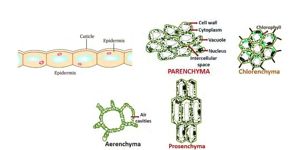
Figure: Different types of parenchyma
Parenchyma is the most common simple permanent tissue. It is a group of living cells that are oval or spherical, with thin cellulose walls, a nucleus, cytoplasm and vacuole. The cells are loosely arranged with intercellular spaces. It is found in the soft parts of the plant and mainly stores food.
Modifications of parenchyma
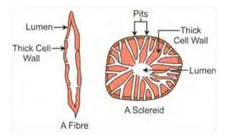
Figure: Sclerenchyma, a fibre and a sclereid
| Fibre | Sclereid |
|---|---|
| Much elongated, narrow, spindle-shaped cell. | Isodiametric (nearly round) cell, also called stone cell. |
| Ends are tapering (pointed). | Cell is short and may be branched. |
| Walls are thick and lignified, lumen is narrow. | Walls are thicker, with a higher degree of lignification. |
| Of three types: surface fibres, wood fibres and bast fibres. | Makes the protective layer of seeds and hard fruits. |
| Gives mechanical support. Fibres of jute, hemp and flax are used to make commercial products. | Gives hardness. Grittiness of guava and pear is due to sclereids, and the stony shell of coconut and walnut is made of them. |
A complex permanent tissue is a group of different kinds of cells that work together as one unit to do a specific function. The cells differ in origin, shape, size and nature. Because they mainly conduct materials, they are also called conducting tissues or vascular tissues.
Types:
| Xylem | Phloem |
|---|---|
| Also called wood or hadrome. | Also called bast or leptome. |
| Conducts water and minerals. | Conducts prepared organic food. |
| Flow is one way, from root to other parts (upward). | Flow is in both directions. |
| Made of tracheids, vessels, xylem parenchyma and xylem fibres. | Made of sieve tubes, companion cells, phloem parenchyma and phloem fibres. |
| Mostly dead cells (only xylem parenchyma is living). | All cells are living except phloem fibres. |
| Gives mechanical strength. | Does not give much mechanical strength. |
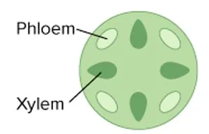
Figure for Q.7
The figure shows a vascular bundle in which phloem (outer side) and xylem (inner side) lie together on the same radius, and there is no strip of cambium between them.
b) Name the type of vascular bundle?
c) Which type of plants have such vascular bundles in stem?
d) Is secondary growth possible in such a vascular bundle? Why?
A transverse section of a young dicot root shows these features.
| Dicot root | Monocot root |
|---|---|
| Vascular bundles are few: 2 to 6 (diarch to hexarch). | Vascular bundles are many: more than 6 (polyarch). |
| Pith is very small or absent. | Pith is large and well developed. |
| Conjunctive tissue is parenchyma and it takes part in forming cambium. | Conjunctive tissue is parenchyma and it may become sclerenchyma in old roots. |
| Pericycle gives rise to lateral roots and vascular cambium. | Pericycle gives rise to lateral roots only. |
| Cambium forms later, so secondary growth takes place. | Cambium is absent, so there is no secondary growth. |
| Cortex is made of parenchyma only. | Cortex of old roots may have a sclerenchymatous exodermis. |
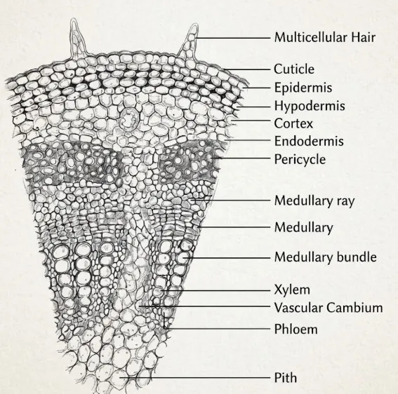
Figure for Q.10
The figure shows a transverse section of a stem. It has an epidermis with hairs, a hypodermis, a cortex, an endodermis, a pericycle, vascular bundles in a ring with cambium in each bundle, medullary rays and a central pith.
a) Name the given figure.
b) What is hypodermis made up of?
c) Write about its vascular bundles.
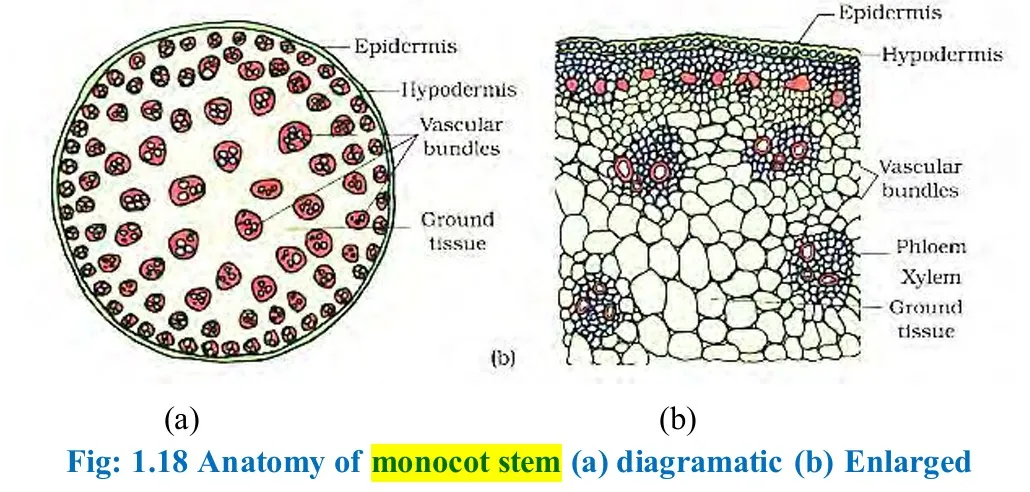
Figure: Plant anatomy of a monocot stem, T.S.
A transverse section of a monocot stem is drawn as below. Its main parts are listed under the diagram.
| Basis | Dicot leaf | Monocot leaf |
|---|---|---|
| Stomatal distribution | Many stomata on the lower epidermis, and few or none on the upper epidermis. | Stomata are present on both upper and lower epidermis, almost equally. |
| Nature of mesophyll tissue | Differentiated into palisade layer (upper) and spongy layer (lower). | Not differentiated into palisade and spongy layers. It is homogeneous. |
| Shape of guard cells | Kidney (bean) shaped. | Dumb-bell shaped. |
| Bundle sheath extension | Made of collenchyma cells. | Made of dead sclerenchyma cells. |
| Bulliform cells | Absent. | Present in the upper epidermis of some monocots (maize). They help in rolling of the leaf. |
| Vascular bundles | Of different sizes, arranged in a net (reticulate venation), conjoint, collateral and closed. | Of different sizes, parallel to each other (parallel venation), conjoint, collateral and closed. |
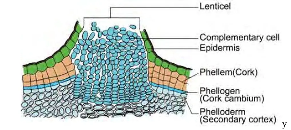
Figure: Anatomy of periderm (phellem, phellogen and phelloderm)
Periderm is the protective tissue formed in the stem and root of dicots and gymnosperms during secondary growth. It replaces the epidermis, which breaks because of the increase in thickness. It is made of three layers, formed by the activity of cork cambium:
At some places the phellogen forms loosely arranged complementary cells instead of cork. They rupture the epidermis to form lenticels, the open pores for gas exchange.
| Early wood (spring wood) | Late wood (autumn wood) |
|---|---|
| Formed in spring, when the cambium is more active. | Formed in dry summer or cold winter, when the cambium is less active. |
| Vessels are more and have wider cavities. | Vessels are fewer and have narrow cavities. |
| Wood is lighter in colour and less dense. | Wood is darker in colour and denser. |
| The band is broad and forms the major part of the ring. | The band is narrow and thin. |
Long Answer Questions
A tissue is a group of cells with a common origin that carry out a specific function. In a plant, tissues build the organs (root, stem, leaf), and the order of organisation is cell, tissue, organ, organ system, organism. Plant tissues can be studied under the microscope by cutting a thin section of the plant part.
Plant tissues are classified on the basis of the power of cell division into meristematic tissue and permanent tissue. The flow chart is shown below.
Figure: Plant anatomy flow chart showing the types of plant tissues
A simple permanent tissue is made of similar cells that have a common origin and do the same work. It is of three types.
1. Parenchyma
Figure: Different types of parenchyma
2. Collenchyma
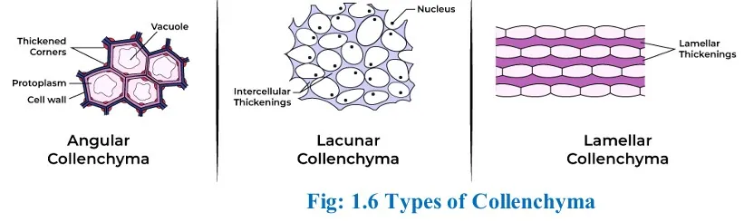
Figure: Collenchyma (plant anatomy of simple permanent tissue)
3. Sclerenchyma
Figure: Sclerenchyma, a fibre and a sclereid
A vascular bundle is a strand of the vascular system of the plant. It is made of xylem and phloem, and cambium may or may not be present. It can be seen under the microscope in any plant part during primary growth.
On the basis of arrangement of xylem and phloem, vascular bundles are of three types.
1. Radial type. Xylem and phloem lie at alternate positions on different radii. It is found in roots.
Figure: Radial vascular bundle (xylem and phloem on different, alternating radii)
2. Conjoint type. Xylem and phloem are attached together as a single bundle. It is of three types.
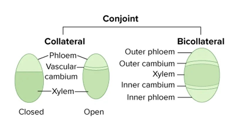
Figure: Conjoint type of vascular bundle (collateral and bicollateral)
3. Concentric type. One of xylem and phloem is completely surrounded by the other. It is of two types.
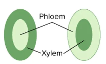
Figure: Concentric type of vascular bundle (amphicribral and amphivasal)
The transverse section of a typical monocot root (for example maize) shows the following tissues.
Figure: Plant anatomy of a monocot root, T.S. (labelled)
Three diagnostic features of the monocot root
Secondary growth is the increase in the thickness (girth) of the root, stem and branches. It is brought about by the lateral meristems, the vascular cambium and the cork cambium. It takes place mainly in dicots and gymnosperms, and not in monocots, pteridophytes and leaves.
Growth of stele (activity of the vascular cambium)
The widening of the stele by the vascular cambium is called intra-stelar secondary growth. It takes place in the following steps.
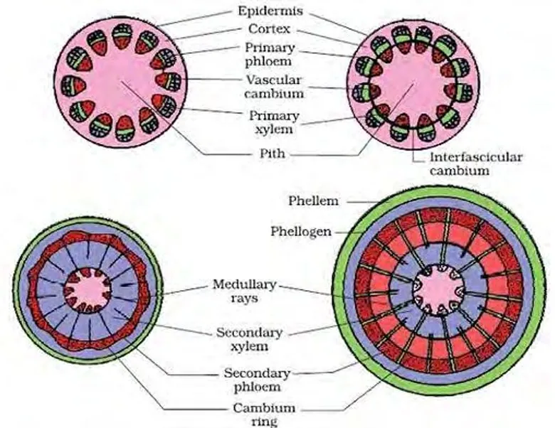
Figure: Growth of the stele during secondary growth in a dicot stem
Extra-stelar secondary growth. The increase in girth ruptures the epidermis and cortex. The outer cortical cells form a cork cambium (phellogen), which makes the periderm (phellem, phellogen and phelloderm) and lenticels. So secondary growth in a dicot stem forms wood in the centre and periderm at the periphery.
Significance: it gives timber, makes the stem strong enough for the tree to grow tall, gives the age of the tree through annual rings, and the corky bark protects the inner tissues.
4. Plant Anatomy Notes PDF
PDF Notes 1
PDF Notes 2
5. Plant Anatomy: Frequently Asked Questions
What is plant anatomy? (Plant anatomy notes)
What are the types of plant tissues?
What is the difference between xylem and phloem?
What are the types of vascular bundles?
What is the difference between dicot and monocot root, stem and leaf?
What is secondary growth in a dicot stem?
Where can I get plant anatomy pdf, plant anatomy notes pdf, plant anatomy ppt and plant anatomy book?
Where can I get Class 12 Biology Book PDF Nepal and Class 12 Biology Book PDF Asmita Publication?
Where can I get Class 12 biology notes, Class 12 biology pdf and Class 12 biology notes pdf for chapter 1?
Where can I get Class 12 Biology notes NEB (Nepal) in the style of Note Mandir?
What topics are included in Unit 1 Plant Anatomy of NEB Class 12 Biology?
Students also search for: